|
Search Term: " deficis "
The Ultimate Guide to Cellular Longevity: NAD+, Methylation, and Senolytics   
Date:
September 10, 2026 10:57 AM
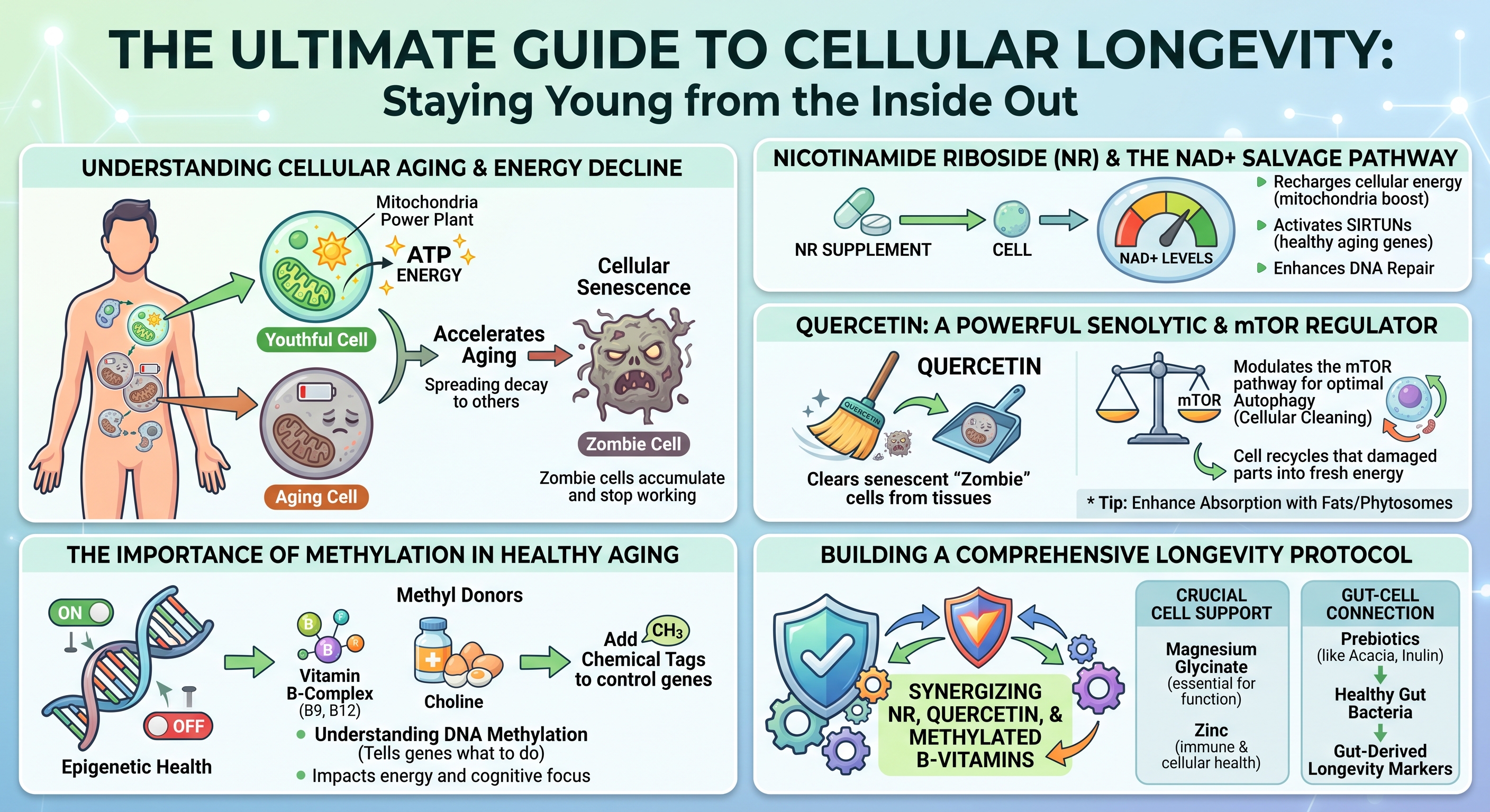
Introduction: Understanding Cellular Aging and Energy DeclineBiological aging represents a progressive decline in cellular maintenance, structural repair, and energy generation. Over decades, tissues experience an attrition of functional reserves, compromised stress resilience, and persistent low-grade systemic inflammation. At the cellular scale, biological degeneration is driven by a failure to generate bioenergetic fuel, repair genetic code, and clear metabolic waste.Cellular aging is characterized by interconnected biological disruptions known as the hallmarks of aging. These encompass genomic instability, epigenetic alterations, mitochondrial decay, loss of proteostasis, and cellular senescence. Rather than operating as isolated occurrences, these phenomena establish a self-reinforcing degenerative cycle: declining cellular power generation impairs enzymatic genetic repair, promoting the accumulation of damaged cells that enter irreversible growth arrest and poison surrounding healthy tissues. Mitigating cellular aging requires examining how microscopic bioenergetic pathways deteriorate and evaluating how targeted nutritional and biochemical interventions can restore cellular homeostasis. The Role of Mitochondria and ATP ProductionEvery biological function - from muscular contraction to continuous DNA replication - depends on adenosine triphosphate (ATP), the primary biochemical energy currency of living systems. Cells produce the vast majority of this energy within mitochondria through oxidative phosphorylation. Within these specialized organelles, metabolic intermediates derived from dietary carbohydrates and lipids donate high-energy electrons to the electron transport chain. The flow of these electrons across protein complexes establishes an electrochemical proton gradient across the inner mitochondrial membrane, driving ATP synthase to manufacture ATP.A youthful cell functions like an efficient municipal power grid, dynamically matching energetic demands with immediate ATP output. However, as biological aging progresses, mitochondrial efficiency declines. The electron transport chain becomes structurally leaky, inadvertently shedding electrons that react with ambient molecular oxygen to produce reactive oxygen species (ROS). While regulated levels of ROS participate in vital intracellular signaling, chronic excess induces widespread oxidative stress. Mitochondria are exceptionally vulnerable to this oxidative burden because they carry their own circular genetic material, known as mitochondrial DNA (mtDNA). Unlike nuclear DNA, mtDNA lacks the protective shielding of histone proteins and possesses rudimentary repair systems. As a result, mtDNA sustains cumulative oxidative damage, encoding increasingly defective electron transport chain proteins. This dynamic generates a bioenergetic deficit: degraded mitochondria synthesize progressively less ATP while emitting greater volumes of damaging free radicals. Deprived of optimal ATP reserves, cells lack the energy necessary to drive vital enzymatic repair cascades, accelerating structural degeneration and functional exhaustion. How Cellular Senescence Accelerates the Aging ProcessWhen healthy cells confront critical physiological damage - such as severe telomere attrition, persistent DNA double-strand breaks, or oxidative stress - they activate protective cell cycle arrest pathways governed primarily by the p53/p21^CIP1 and p16^INK4a/Rb molecular checkpoints. This defensive shutdown, termed cellular senescence, permanently prevents the replication of potentially premalignant or mutated cells.Senescent cells, colloquially known as "zombie cells," enter a state of permanent growth arrest while actively resisting programmed cell death (apoptosis). Over time, these cells accumulate within adipose depots, skeletal muscle, the vascular endothelium, and major organs, largely because immune surveillance and clearance pathways simultaneously lose functional efficiency. The systemic danger of senescent cells stems from their secretome. Rather than remaining biologically inert, senescent cells develop a hyperactive secretory state termed the Senescence-Associated Secretory Phenotype (SASP). The SASP is a destructive mixture of pro-inflammatory cytokines, chemokines, extracellular matrix-degrading matrix metalloproteinases (MMPs), and reactive oxygen species. Through this toxic secretome, even a small burden of senescent cells can impair whole-tissue architecture. SASP factors degrade surrounding structural proteins, induce insulin resistance in neighboring metabolic cells, and biochemically force adjacent healthy cells into secondary senescence. This persistent paracrine signaling fuels chronic, sterile, low-grade systemic inflammation, termed "inflammaging," which accelerates systemic tissue degeneration and elevates susceptibility to degenerative age-related pathologies. Nicotinamide Riboside (NR) and the NAD+ Salvage PathwayThe Biochemistry of NAD+ Depletion Over TimeNicotinamide adenine dinucleotide (NAD+) is an indispensable coenzyme present in every living cell. NAD+ fulfills a dual biological mandate: it serves as a central redox cofactor that shuttles electrons between cellular metabolic reactions, and it functions as an obligatory consumable substrate for regulatory enzymes that preserve cellular viability. In its redox capacity, NAD+ accepts electrons to form NADH during glycolysis, the tricarboxylic acid (TCA) cycle, and fatty acid beta-oxidation, subsequently donating those electrons to Complex I of the respiratory chain to power ATP synthesis.
The primary enzymatic driver of age-related NAD+ destruction is CD38, a membrane-bound glycohydrolase expressed on immune cells that is upregulated in response to chronic SASP exposure. Concurrently, lifelong genotoxic damage causes persistent activation of Poly(ADP-ribose) polymerase 1 (PARP-1), an enzyme that cleaves the glycosidic bonds of NAD+ to assemble branched poly(ADP-ribose) chains at DNA lesion sites. Because PARP-1 consumes NAD+ without directly recycling the molecule, chronic DNA damage depletes intracellular NAD+ pools, impairing bioenergetics and limiting sirtuin activity. How NR Efficiently Boosts Cellular NAD+ LevelsThe mammalian body maintains its NAD+ supply through three distinct biosynthetic routes: the de novo pathway from dietary L-tryptophan, the Preiss-Handler pathway from nicotinic acid (niacin), and the NAD+ Salvage Pathway. The de novo pathway requires substantial energy expenditure, consuming roughly sixty milligrams of dietary tryptophan to yield a single milligram of NAD+. The Preiss-Handler pathway, while effective, can induce cutaneous prostaglandin-mediated flushing at therapeutic intakes. Consequently, the salvage pathway serves as the primary mechanism for maintaining intracellular NAD+ pools.The salvage pathway recycles the breakdown product nicotinamide (NAM), which is released whenever NAD+-consuming enzymes execute their functions. Under normal conditions, cells convert free nicotinamide into nicotinamide mononucleotide (NMN) via the rate-limiting enzyme nicotinamide phosphoribosyltransferase (NAMPT), after which NMN adenylyltransferases (NMNAT1–3) complete the conversion into NAD+. However, NAMPT expression declines with advancing age, chronic inflammation, and metabolic stress, limiting the recycling capacity of the cell. Nicotinamide Riboside (NR) is a naturally occurring pyridine nucleoside that bypasses this enzymatic bottleneck. Upon cellular entry via equilibrative nucleoside transporters, NR is directly phosphorylated into NMN by nicotinamide riboside kinases (NRK1 and NRK2) using a single molecule of ATP. Because the NRK pathway remains intact and robust across the lifespan, NR provides an efficient alternative entry point into the NAD+ salvage cascade. Clinical evaluations in humans confirm the safety, bioavailability, and pharmacokinetics of oral NR supplementation. Randomized, double-blind, placebo-controlled trials reveal that oral NR chloride produces dose-dependent increases in steady-state whole blood NAD+ concentrations. Dosing regimens of 100 mg, 300 mg, and 1,000 mg daily elevate blood NAD+ levels by approximately 22%, 51%, and up to 142%, respectively, within two weeks of administration, maintaining these elevations throughout continuous use. High-resolution metabolomic analyses also demonstrate parallel elevations in nicotinic acid adenine dinucleotide (NAAD), establishing it as a reliable biomarker of active intracellular NAD+ synthesis without hepatic or systemic toxicity. Sirtuin Activation and DNA Repair MechanismsReplenishing intracellular NAD+ supports functions beyond mitochondrial ATP generation. NAD+ functions as an obligatory cofactor for sirtuins (SIRT1 through SIRT7), a family of class III histone and non-histone protein deacetylases that regulate stress resilience, metabolic homeostasis, and cell survival. Sirtuins couple the removal of acetyl groups from target lysine residues to the stoichiometric cleavage of NAD+, producing nicotinamide and O-acetyl-ADP-ribose. In states of NAD+ deficiency, sirtuin enzymes remain inactive regardless of cellular demand.In the nucleus, SIRT1 coordinates defense against cellular decline. When activated by restored NAD+ levels, SIRT1 deacetylates peroxisome proliferator-activated receptor-gamma coactivator 1-alpha (PGC-1a), the master transcriptional coactivator of mitochondrial biogenesis. This deacetylation stimulates mitochondrial replication and assembly, expanding functional respiratory capacity. Concurrently, SIRT1 deacetylates the p65 subunit of nuclear factor-kappa B (NF-kB), suppressing the transcription of pro-inflammatory cytokines. In the mitochondria, SIRT3 utilizes NAD+ to deacetylate metabolic enzymes and superoxide dismutase 2 (SOD2), enhancing the organelle's capacity to neutralize reactive oxygen species. At the same time, cellular NAD+ levels directly regulate genomic integrity through PARP-1. When genotoxic stress or oxidative damage induces single- or double-strand DNA breaks, PARP-1 binds to the damaged termini using its zinc-finger domains. Bound PARP-1 hydrolyzes NAD+ to synthesize extensive, negatively charged poly(ADP-ribose) polymers on itself and adjacent histones. This modification relaxes chromatin architecture and establishes an electrostatic scaffold that recruits base excision repair and homologous recombination complexes. Recent discoveries demonstrate close crosstalk between sirtuins and PARP-1 during DNA repair. PARP-1 recruits SIRT1 to double-strand breaks, where SIRT1 deacetylates the chromatin-remodeling ATPase BRG1 to displace nucleosomes and facilitate homologous recombination. However, because PARP-1 and SIRT1 draw from the same intracellular NAD+ pool, severe NAD+ depletion forces a biological compromise: PARP-1 consumes the scarce remaining cofactor to address DNA damage, leaving sirtuins deactivated. Restoring NAD+ via NR prevents this deficit, enabling concurrent genomic repair and sirtuin-mediated metabolic defense. Quercetin: A Powerful Senolytic and mTOR RegulatorClearing Senescent "Zombie" Cells from TissuesThe accumulation of senescent cells has driven interest in senolytics: molecules that selectively eliminate senescent cells while sparing healthy, non-senescent populations. Senolytic agents exploit a specific vulnerability in senescent cells. Because senescent cells produce cytotoxic, pro-inflammatory SASP factors that would normally induce their own death, they become dependent on upregulated Senescent Cell Anti-Apoptotic Pathways (SCAPs) to survive. The SCAP network involves anti-apoptotic proteins (such as BCL-2 and BCL-xL), the PI3K/Akt kinase cascade, and cyclin-dependent kinase inhibitors.Quercetin is a polyphenolic flavonoid found in capers, red onions, apples, and the flower buds of Sophora japonica. Beyond its classical antioxidant properties, quercetin functions as a senolytic compound that exerts multi-target inhibitory effects across the SCAP network. By inhibiting the upstream PI3K/Akt survival axis and downregulating anti-apoptotic defenses, quercetin disrupts the signaling that protects senescent cells from intrinsic apoptosis. Deprived of these survival signals, senescent cells undergo programmed cell death. Preclinical studies demonstrate that senolytic protocols utilizing quercetin - often combined with the tyrosine kinase inhibitor dasatinib - reduce senescent cell burden across multiple tissues. This targeted clearance lowers circulating SASP factors, attenuates tissue fibrosis, restores endothelial reactivity, and improves functional health span. By removing senescent cells, quercetin mitigates the primary driver of chronic, low-grade inflammaging. Modulating the mTOR Pathway for Optimal AutophagyThe mechanistic Target of Rapamycin (mTOR) is an evolutionarily conserved serine/threonine protein kinase that coordinates cellular metabolism by balancing anabolic growth with catabolic recycling. Operating within two multiprotein complexes - mTORC1 and mTORC2 - the mTOR pathway integrates signals from amino acids, growth factors, and intracellular energy levels. In nutrient-rich environments, mTORC1 promotes protein synthesis, lipogenesis, and cellular growth, while suppressing catabolic breakdown. Conversely, nutrient scarcity downregulates mTORC1, activating autophagy.Autophagy is an intracellular degradation system that packages damaged organelles, misfolded protein aggregates, and biological debris into double-membraned autophagosomes for lysosomal degradation and recycling. A specialized branch of this pathway, mitophagy, selectively targets and clears damaged mitochondria. In modern metabolic conditions characterized by continuous caloric intake, mTORC1 can remain persistently active. This persistent signaling suppresses autophagy, causing damaged organelles and toxic aggregates to accumulate within tissues. Quercetin functions as a natural modulator of mTOR signaling. By inhibiting upstream PI3K/Akt signaling and activating intracellular energy sensors, quercetin attenuates overactive mTORC1, mimicking the metabolic effects of caloric restriction. This down-regulation relieves inhibition on the ULK1 autophagy initiation complex, stimulating both general autophagy and mitophagy. As autophagy proceeds, cells clear protein aggregates and eliminate damaged mitochondria, supporting cellular longevity and proteostasis. Enhancing Absorption: Phytosomes and Dietary FatsDespite the biological activities of quercetin identified in experimental models, its clinical translation has historically been limited by poor oral bioavailability. Raw quercetin aglycone is a crystalline, hydrophobic polyphenol with poor solubility in water and gastrointestinal fluids. When ingested in unformulated powder forms, quercetin molecules aggregate in the gut lumen, resisting dissolution and passive absorption. Consequently, the vast majority of an unformulated dose passes into the colon unabsorbed, where it undergoes microbial degradation without reaching meaningful systemic concentrations.To address these pharmacokinetic limitations, advanced delivery systems such as phytosomes were engineered. A phytosome is a 100% food-grade molecular complex where individual polyphenolic molecules are bound to dietary phospholipids, typically sunflower-derived phosphatidylcholine. Unlike a classical liposome - which encapsulates water-soluble compounds inside an aqueous core enclosed by a lipid bilayer - a phytosome forms an amphiphilic complex at the molecular level. The polar head of the phosphatidylcholine molecule forms hydrogen bonds with the hydroxyl groups of the quercetin molecule, while its lipophilic fatty acid tails extend outward. This structural arrangement shields the polar regions of the flavonoid, creating a lipid-compatible complex that integrates smoothly into the intestinal mucosa.
The Importance of Methylation in Healthy AgingVitamin B-Complex and Choline as Essential Methyl DonorsMethylation is an essential biochemical process occurring billions of times each second across all human tissues. It involves the transfer of a single-carbon unit - a methyl group consisting of one carbon atom bound to three hydrogen atoms - (CH3) - from a donor molecule to diverse recipients, including DNA, RNA, structural proteins, neurotransmitters, and membrane phospholipids. This transfer of one-carbon units is coordinated by the methionine-homocysteine cycle, which sustains genetic stability, detoxification pathways, and cellular repair.At the center of this pathway sits S-adenosylmethionine (SAM), the universal methyl donor in human biology. When a methyltransferase enzyme transfers a methyl group from SAM to an acceptor molecule, SAM is converted into S-adenosylhomocysteine (SAH). SAH functions as a potent competitive inhibitor of intracellular methyltransferases. To maintain functional methylation, SAH is rapidly hydrolyzed into homocysteine, a sulfur-containing amino acid that must be remethylated or cleared through transsulfuration. Homocysteine clearance proceeds through two distinct remethylation pathways. The primary route operates across most tissues via the enzyme methionine synthase, which requires vitamin B12 in its active methylcobalamin form. Methionine synthase transfers a methyl group from 5-methyltetrahydrofolate (5-MTHF, the active form of folate) to homocysteine, regenerating methionine. The ongoing production of 5-MTHF depends on the enzyme methylenetetrahydrofolate reductase (MTHFR), which utilizes riboflavin (vitamin B2) as a cofactor. Alternatively, excess homocysteine can be routed into the transsulfuration pathway by vitamin B6 (as pyridoxal-5'-phosphate) to synthesize cystathionine, cysteine, and ultimately the antioxidant glutathione. A secondary remethylation pathway, active predominantly in hepatic and renal tissues, bypasses folate entirely. In this route, dietary choline is oxidized to betaine (trimethylglycine or TMG). The enzyme betaine-homocysteine S-methyltransferase (BHMT) then transfers a methyl group from betaine directly to homocysteine, yielding methionine and dimethylglycine. When dietary intake of active B-vitamins or choline is insufficient, or when genetic variations like MTHFR polymorphisms reduce pathway flux, the methylation cycle slows. Homocysteine accumulates in circulation, promoting vascular and neurological inflammation, while SAM reserves decline, restricting cellular methylation capacity. Understanding DNA Methylation and Epigenetic HealthEvery somatic cell in an organism carries an identical genetic code. Cellular differentiation and tissue-specific functions are governed by the epigenome: a regulatory layer of chemical modifications that dictates gene expression without altering underlying DNA sequences. DNA methylation represents the primary and most stable epigenetic modification. In this process, DNA methyltransferase (DNMT) enzymes utilize methyl groups donated by SAM to add a methyl tag to cytosine bases adjacent to guanine residues, forming 5-methylcytosine within CpG dinucleotide sites.Under physiological conditions, DNA methylation maintains genomic stability and coordinates transcription. Methylation of promoter regions condenses chromatin, repressing transposable elements and silencing genes inappropriate for a given cell type. Conversely, hypomethylated promoters maintain an open chromatin state, allowing transcription factors to bind and initiate gene expression. During biological aging, this epigenetic landscape undergoes progressive dysregulation, a phenomenon termed "epigenetic drift". Aging cells experience global hypomethylation alongside focal hypermethylation of specific gene promoters. Global loss of methyl tags destabilizes the genome, activating retrotransposons and pro-inflammatory pathways. Simultaneously, hypermethylation at targeted promoter sites silences critical tumor suppressor genes and DNA repair complexes. This systematic change in DNA methylation patterns is consistent across populations, allowing researchers to develop molecular "epigenetic clocks". Algorithms such as the Horvath clock, PhenoAge, and GrimAge quantify biological age by profiling the methylation status of specific CpG sites across the genome. These clocks assess whether individuals are aging faster or slower than their chronological years. Ensuring a steady supply of methyl donors and preventing unnecessary SAM depletion supports DNMT activity, maintaining epigenetic patterns and genomic stability. How the Methylation Cycle Impacts Energy and Cognitive FocusBeyond long-term epigenetic regulation, the methylation cycle directly modulates immediate biochemical processes that govern daily energy, neurotransmission, and cognitive focus. Compromised methylation capacity frequently manifests as cognitive slowing, executive fatigue, and reduced physical stamina.A major consumer of methyl reserves is the endogenous synthesis of creatine. Approximately 40% of all SAM-derived methyl groups in the human body are utilized by guanidinoacetate N-methyltransferase (GAMT) in the liver to synthesize creatine. Creatine then translocates to the brain and skeletal muscle, where it is phosphorylated into phosphocreatine. Phosphocreatine functions as a rapid energy buffer, donating a high-energy phosphate group to regenerate ADP into ATP in milliseconds during demanding physical or cognitive tasks. When methyl donor availability falls, endogenous creatine synthesis drops, depleting phosphocreatine reserves and increasing susceptibility to neuromuscular and cognitive fatigue. Methylation is equally central to central nervous system architecture. SAM provides methyl groups to convert phosphatidylethanolamine into phosphatidylcholine, the predominant phospholipid comprising neuronal cell membranes and the myelin sheaths that insulate axons. Intact myelin preserves rapid action potential conduction throughout the nervous system. Furthermore, free choline derived from this pathway is the direct precursor to acetylcholine, the neurotransmitter required for attention, working memory, and learning. The methylation cycle also governs monoamine neurotransmitter metabolism. SAM is required for the synthesis of adrenaline (epinephrine) from noradrenaline, while catechol-O-methyltransferase (COMT) relies on SAM to degrade dopamine and norepinephrine within the prefrontal cortex. Sluggish methylation disrupts this balance, contributing to cognitive fatigue, mood variability, and impaired mental performance. Building a Comprehensive Longevity ProtocolSynergizing NR, Quercetin, and Methylated B-VitaminsLongevity supplementation often falters when single molecules are administered in isolation, ignoring interconnected metabolic pathways. Designing an effective cellular longevity protocol requires combining complementary mechanisms that reinforce one another while preventing secondary metabolic deficits. The combination of Nicotinamide Riboside, Quercetin Phytosome, and Methylated B-Vitamins illustrates this multi-target synergy.This synergy is grounded in the direct biochemical intersection between the NAD+ salvage pathway and the methylation cycle. When high-dose NR is supplemented to boost systemic NAD+, sirtuins and PARP enzymes consume the newly synthesized cofactor, generating substantial quantities of free nicotinamide (NAM). This intracellular nicotinamide faces two primary metabolic fates: it can be recycled back into NAD+ through the NAMPT-dependent salvage loop, or it can be cleared via methylation. When the influx of nicotinamide exceeds salvage recycling capacity, the excess is cleared to avoid feedback inhibition of sirtuin enzymes. To accomplish this, the enzyme nicotinamide N-methyltransferase (NNMT) transfers a methyl group from SAM directly onto nicotinamide, forming 1-methylnicotinamide (1-MNA/MNAM), which is subsequently excreted in urine. Prolonged, high-dose precursor administration without nutritional methyl support can elevate NNMT flux, depleting intracellular SAM reserves. As methyl groups are consumed clearing nicotinamide, the cellular SAM-to-SAH ratio falls, which can elevate circulating homocysteine and reduce methyl availability for DNA methylation and neurotransmitter synthesis. Co-administering a fully methylated B-complex alongside choline or betaine addresses this potential bottleneck. Providing active methyl donors (such as 5-MTHF, methylcobalamin, and betaine) maintains the one-carbon donor pool. Even during increased NNMT activity, SAM pools remain stable, protecting DNA methylation fidelity and maintaining homocysteine within safe parameters. Quercetin reinforces this protocol through complementary mechanisms. By clearing senescent cells and reducing SASP-mediated inflammation, quercetin downregulates CD38, the primary enzyme responsible for age-related NAD+ degradation. Suppressing CD38 prevents unnecessary breakdown of newly synthesized NAD+, enhancing the efficiency of NR supplementation. Furthermore, while NR provides the NAD+ necessary to activate SIRT1-driven mitochondrial biogenesis, quercetin concurrently modulates mTORC1 to stimulate autophagy. This coordinated action ensures that newly generated mitochondria operate in an environment cleared of proteotoxic cellular debris. The Crucial Role of Magnesium Glycinate and Zinc in Cellular FunctionLongevity protocols require essential mineral cofactors to function efficiently. Without adequate divalent minerals acting as enzymatic cofactors and structural stabilizers, metabolic longevity pathways cannot operate at full capacity. Among these, magnesium and zinc are required for cellular repair, genomic stability, and energy production.Magnesium serves as an obligatory cofactor in over 300 enzymatic reactions, primarily through its interaction with ATP. In biological systems, ATP exists predominantly as a chelate with a divalent magnesium ion, forming biologically active Mg2+ -ATP. Every enzymatic reaction that synthesizes, transfers, or consumes cellular energy - including the enzymes of the NAD+ salvage pathway (NRK and NMNAT) and DNA polymerases - strictly requires Mg2+ -ATP as its substrate. Magnesium deficiency impairs these phosphorylation reactions, reducing the cellular utilization of NAD+ precursors. Additionally, magnesium is an essential cofactor for the enzymes that activate dietary B-vitamins into their active forms. Supplying magnesium as magnesium glycinate provides high gastrointestinal bioavailability, minimal laxative effect, and yields glycine to support inhibitory neurotransmission and restful sleep. Zinc serves as a vital structural component for more than 3,000 human transcription factors and enzymatic proteins. Its most prominent structural role in longevity occurs within zinc-finger motifs. These are specialized protein conformations stabilized by a zinc ion coordinated to cysteine and histidine residues. The DNA damage sensor PARP-1 utilizes three zinc-finger domains to identify, track, and physically bind to single- and double-strand DNA breaks. Without adequate intracellular zinc, PARP-1 cannot properly assemble or dock onto damaged chromosomes, impairing DNA repair and increasing genomic instability. Zinc is also an obligatory structural component of copper/zinc superoxide dismutase (Cu/Zn-SOD or SOD1), the primary cytosolic antioxidant enzyme that dismutates superoxide radicals into hydrogen peroxide, protecting mitochondrial membranes and nuclear DNA from premature senescence. Integrating Prebiotics (like Acacia and Inulin) for Gut-Derived Longevity MarkersA comprehensive cellular longevity framework must extend beyond somatic tissues to encompass the gut microbiome. The intestinal microbiome functions as a central regulator of systemic inflammatory tone, immune development, and metabolic signaling. Age-associated dysbiosis - characterized by the loss of beneficial commensals and an overgrowth of pathobionts - frequently leads to breakdown of the intestinal barrier.The gut epithelium consists of a single-cell monolayer sealed by tight junction proteins, including zonula occludens-1 (ZO-1), occludin, and claudins. When this physical barrier is disrupted by poor dietary fiber intake or dysbiosis, gut permeability increases. This allows lipopolysaccharide (LPS), a component of the outer membrane of Gram-negative bacteria, to enter the portal and systemic circulation. The resulting "metabolic endotoxemia" activates Toll-like receptor 4 (TLR4) on immune cells, inducing NF-kB and systemic pro-inflammatory cytokine production. This persistent gut-derived inflammation exacerbates the SASP, accelerates tissue senescence, upregulates CD38, and drains systemic NAD+ reserves.
These short-chain fatty acids, particularly butyrate, exert direct protective effects on systemic longevity. Butyrate provides the primary metabolic fuel for colonic epithelial cells, supplying more than 70% of their baseline energy needs and supporting mitochondrial function within colonocytes. Furthermore, SCFAs upregulate the expression of epithelial tight junction proteins (ZO-1, occludin, and claudin-1), restoring intestinal barrier integrity and preventing the translocation of inflammatory LPS into systemic circulation. Systemically absorbed butyrate also functions as an endogenous histone deacetylase (HDAC) inhibitor, suppressing pro-inflammatory gene expression and supporting regulatory T cell (T_reg) development. Reducing metabolic endotoxemia dampens systemic inflammation, protecting vascular function and preventing premature NAD+ depletion. Conclusion: The Integrated Cellular Longevity MatrixCellular longevity is achieved not by addressing isolated biomarkers in isolation, but by systematically supporting interconnected biological pathways. As bioenergetic capacity declines, cellular senescence accelerates, epigenetic patterns degrade, and gut barrier integrity weakens. A comprehensive approach addresses these biological vulnerabilities simultaneously.
Quercetin Phytosome clears senescent cells and modulates mTORC1, stimulating autophagy while dampening the inflammatory SASP cascade that accelerates CD38-mediated NAD+ destruction. Methylated B-vitamins, active folate, and choline replenish SAM reserves, balancing the methyl requirements of NNMT-mediated nicotinamide clearance, preserving epigenetic DNA methylation, and maintaining neurotransmitter production. Magnesium glycinate and zinc provide the structural and catalytic foundation required for ATP utilization, B-vitamin activation, and PARP-1 zinc-finger DNA repair docking. Finally, prebiotic fibers generate short-chain fatty acids like butyrate, reinforcing the intestinal barrier and preventing metabolic endotoxemia from fueling systemic inflammation. By coordinating energy replenishment, cellular waste clearance, epigenetic maintenance, and the suppression of systemic inflammation, this unified approach directly addresses the underlying drivers of cellular aging to support long-term physiological vitality.
--
(https://vitanetonline.com:443/forums/Index.cfm?CFApp=1&Message_ID=6650) Is there anything else DLPA can do be sides Pain management and Mood? 
Date:
May 22, 2026 04:29 PM
Here is what else DLPA can do:
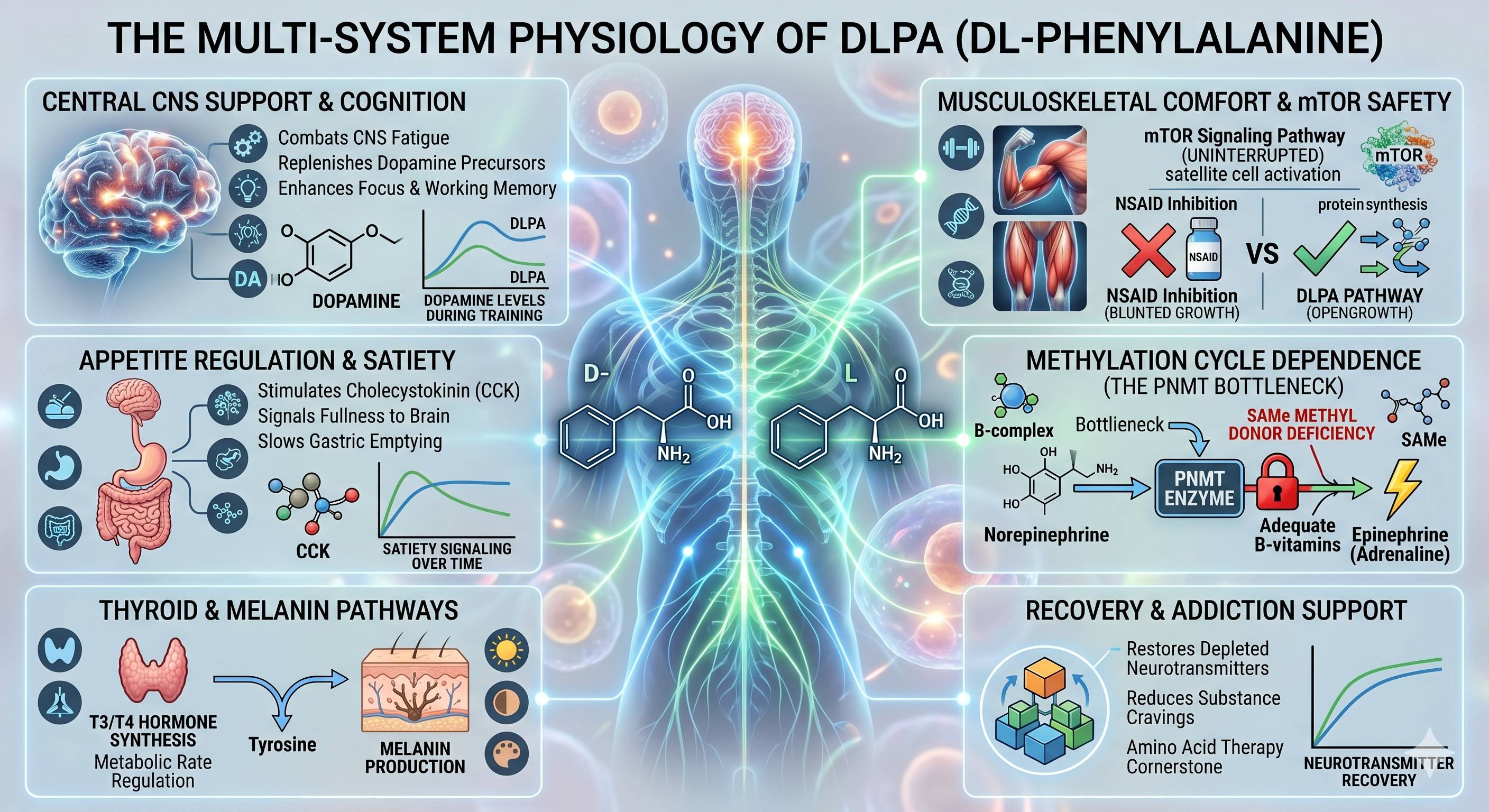
When diving deeper into the physiological applications of DLPA, there are several advanced, highly specific mechanisms that make it particularly interesting, especially regarding athletic performance, metabolic pathways, and cellular recovery.
Here are a few of the more complex, nuanced roles of DLPA in the body:
By supplementing with L-phenylalanine, the brain has a steady supply of precursor material to maintain dopamine synthesis. This helps delay the onset of CNS fatigue, allowing for sustained mental drive, higher intensity, and better neurological output throughout a grueling session.
Because the D-phenylalanine in DLPA does not interfere with inflammation or COX enzymes—working entirely by shielding circulating endorphins—it provides musculoskeletal comfort without compromising the cellular signaling required for muscle repair and growth. It allows the body's natural inflammatory repair mechanisms to run their course uninterrupted.
The enzyme responsible for this final step (PNMT) relies entirely on S-adenosylmethionine (SAMe) to donate a methyl group. If the methylation cycle is sluggish or deficient in necessary cofactors (like methylated B-vitamins), the catecholamine cascade bottlenecks at norepinephrine. Therefore, DLPA's ability to fully support the adrenal axis is directly dependent on the efficiency of the body's methylation pathways.
Would you like to explore how to position this mTOR-safe, CNS-supporting angle into a specific educational breakdown for a retail or blog audience?
Summary:
Beyond its foundational role in chronic pain relief, DLPA serves as a versatile precursor that supports several advanced metabolic, cognitive, and physical functions. Because the L-phenylalanine half converts into tyrosine, it provides the essential raw materials required for thyroid hormone synthesis (T3 and T4), melanin skin pigmentation production, and the release of cholecystokinin (CCK), a gut peptide that signals satiety and regulates appetite. Furthermore, by elevating dopamine and norepinephrine, DLPA enhances executive function, working memory, and focus, making it a valuable asset in nootropic protocols and a supportive tool for rebuilding depleted neurotransmitter stores during substance recovery.
In the context of high-intensity athletic performance, DLPA offers unique physiological advantages for training and recovery. It directly combats central nervous system (CNS) fatigue by replenishing the dopamine depleted during heavy resistance training, thereby maintaining neural drive without interrupting muscle protein synthesis. Unlike traditional NSAIDs, which blunt the acute inflammatory response and stall the mTOR pathway, DLPA provides musculoskeletal comfort solely by shielding endogenous opioids (endorphins and enkephalins) from enzymatic breakdown. This allows the natural muscle-repair mechanisms to run their course uninterrupted while simultaneously prolonging the post-exercise "lifter's high." However, its full efficacy remains dependent on a healthy methylation cycle, as the final conversion of norepinephrine to adrenaline requires adequate methyl donors like SAMe.
(https://vitanetonline.com:443/forums/Index.cfm?CFApp=1&Message_ID=6640)
Date:
January 23, 2018 07:59 AM
There was a study conducted at the Harbin Medical University in China that found that Morin, a flavanoid in guava and osage oranges could lessen inflamation and increase neurological deficits. It also has anti inflamation properties. In labratory mice this flavanoid seemed to reduce inflamation and restored the ability to walk, talk ( which is strange for a labratory mouse.), decreased mental function issues, and decreased loss of balance, and a weakness in the arms and legs. Essentially the study found that the flavanoid morin helped to counter act certain types of strokes, to increase motor functions to the patient. Key Takeaways: "A person who smokes, drinks excessive amounts of alcohol, uses cocaine or methamphetamines, is overweight, or has diabetes is at risk of developing cerebral ischemia." Read more: https://www.naturalnews.com/2018-01-21-a-flavonoid-found-in-guava-and-osage-orange-antioxidant-properties.html
(https://vitanetonline.com:443/forums/Index.cfm?CFApp=1&Message_ID=5501)
Date:
August 12, 2015 08:24 PM
Selenium is nature’s best secret. It is a nutrient that when used in the correct amounts in your food, can make huge and exciting changes. However, the body only needs small amounts of this mineral, but it has huge benefits on one’s health. It protects us against many health problems that are associated with the aging process such as osteoarthritis and cancer. A certain amount of this mineral is required in order to protect the body from deadly viruses and also to aid in healthy thyroid functioning. Selenium deficiency frequently goes untreated simply because it does not show any noticeable symptoms. If the levels of selenium are low, the thyroid will have to work extra hard to manufacture its hormones and the body will have a tough job converting those hormones to a form the cells can use. This is because selenium is a chief ingredient of the molecules necessary for the body to be able to create and use seleno-proteins (thyroid hormones). The initial stages of low selenium levels will reduce the optimal production of the thyroid hormone in the glands. If this continues the thyroid gland becomes inflamed. Eventually, this is what leads to thyroid autoimmune problems Problems that come with low selenium levels can be more serious when the levels of mineral, iodine, is low. Iodine is well known for its key role in thyroid health. Selenium is also important in aiding your body to recycle iodine. Low selenium levels coupled with low iodine levels can lead to thyroid imbalance An enlarged thyroid is known as goiter. This is one of the serious forms of thyroid imbalance. Most individuals diagnosed with goiter usually have low iodine levels, however research has also shown that the patient also have low levels of selenium. Therefore, it is very important to treat both deficits so as to re-establish thyroid stability Doctors readily admit that there is no any specified dose of selenium required for better functioning of the thyroid. The one thing they agree on is that optimizing selenium intake aids in general health. References //www.google.com/url?sa=t&rct=j&q=&esrc=s&source=web&cd=5&cad=rja&uact=8&ved=0CDYQFjAEahUKEwih0urP8KPHAhXHOBQKHfIrC3w&url=http%3A%2F%2Fwww.nlm.nih.gov%2Fmedlineplus%2Fdruginfo%2Fnatural%2F1003.html&ei=8GjLVaGaCsfxUPLXrOAH&usg=AFQjCNHzSWE2P10XlrjScWz9oE6KOYZFdA&bvm=bv.99804247,d.d24 //www.google.com/url?sa=t&rct=j&q=&esrc=s&source=web&cd=9&cad=rja&uact=8&ved=0CE4QFjAIahUKEwih0urP8KPHAhXHOBQKHfIrC3w&url=http%3A%2F%2Fpatient.info%2Fdoctor%2Fselenium&ei=8GjLVaGaCsfxUPLXrOAH&usg=AFQjCNEjLPYH8bzUP0K2eUgEAAR7HIrZAg&bvm=bv.99804247,d.d24
(https://vitanetonline.com:443/forums/Index.cfm?CFApp=1&Message_ID=3192)
Date:
August 04, 2014 08:16 PM
Memory loss occurs when one loses the ability to remember information and events that you would normally be able to recall. It is usually distressing for the person affected and the family members. Gingko Biloba Benefits Ginkgo biloba is effective for memory aid as it improves blood flow to the brain. It can help with memory problems caused by dementia or Alzheimer's disease. It helps to prevent the progression of dementia symptoms. It also helps in the improvement in cognitive function. Taking ginkgo biloba leaf extract by mouth seems to improve thinking skills in elderly people with mild moderate age related memory loss or thinking problems. It improves short-term visual memory and speed of mental processing in non-demented people with age related memory loss and it can improve memory and speed of mental processing in people without memory loss. Ingredients of Gingko Biloba The active ingredients in ginkgo biloba are ginkgolides, bilobalides and flavonoids. It works by delaying the symptoms of dementia. Adding ginkgo biloba supplements may help battle cognitive impairments and memory loss associated with dementia since it encourages the growth and development of neural stem cells. It protects the neurons from oxidative damage by potentially preventing the progression of tissue generation in patients suffering from dementia. Ginkgo biloba extracts promotes growth in brain regions. Due to its benefit of boosting memory, most doctors prescribe ginkgo biloba as the treatment choice for dementia and for age related memory dysfunction. It has been approved for symptomatic treatment of deficits in memory impairment, concentration difficulties and depression from organic brain disease. In conclusion, it is evident that ginkgo biloba extracts helps to stabilize and improve memory loss in several measures of social functioning and cognitive functioning in mixed or vascular dementia patients and Alzheimer's Disease. It might also improve memory and speed of mental processing in people without memory loss.
(https://vitanetonline.com:443/forums/Index.cfm?CFApp=1&Message_ID=3152)
Date:
July 18, 2008 12:36 PM
You can be sure that a supplement has some remarkable therapeutic possibilities when it makes headlines on the five o’clock news. A recent experiment which showed the conditions of rats that had been fed a diet that was totally deficient in vitamin E tested alpha lipoic acid, which caused the rats to return to full health even though no vitamin E was replenished. The results of this test are profound, as giving these rats an alpha lipoic acid caused existing stores of vitamin E that the body was previously unable to use to be regenerated.
Alpha lipoic acid is a vitamin-like antioxidant that has been used in Europe for a long period of time. It has recently emerged as an extremely impressive therapeutic agent that scavenges free-radicals. Recent studies have suggested that it has the ability to stop some degenerative diseases, the oxidative process of aging, and restores the health of diseased organs. Additionally, it has the ability to make up deficits of vitamin E or C and could potentially be one of the best treatments that have emerged for diabetes. Alpha lipoic acid (ALA) is considered to be more potent than vitamins E and C, as well as coenzyme Q10. Unlike other antioxidants, ALA has properties that make it superior because it is able to replace certain nutritional supplements, while potentiating others, and inhibiting tissues from deterioration that is associated with diseases. This supplement is also both fat and water soluble, which allows it to protect lipid and aqueous cell structures.
Alpha lipoic acid is a compound that is synthesized in small amounts in the body, but can also be supplied from food or supplement sources. A vitamin-like substance, it contains sulfur and also plays a crucial role in energy reactions. It can be found in liver, yeast, spinach, organ meats, broccoli, red potatoes, and red meat. When it is orally ingested, alpha lipoic acid is not compromised in the GI tract or the liver.
Numerous studies have been conducted on ALA, all of which confirm its positive effect on metabolic processes, with recent clinical tests supporting its ability to enhance free-radical protection, slow the aging process, and guard against a variety of degenerative diseases. ALA was discovered in the 1930s, where it was originally classified as a vitamin, and later categorized as an essential coenzyme when scientists discovered that it was involved in the energy processes of cell mitochondria. It wasn’t until 1988 that scientists found that it also has powerful antioxidant effects.
Alpha lipoic acid is important because it protects us from free-radicals which are present in a body as a result of the number of toxic substances such as auto exhaust, tobacco smoke, pollution, preservatives, and additives that we are exposed to on a daily basis. These free radicals can actually accelerate the aging process, causing premature tissue breakdown to occur. Additionally, our environment will continue to surround us with these pollutants that create free radicals.
There are things we can do to minimize our health risks, which include exercising, eating nutritiously, and not smoking. However, these measures are rarely enough to decrease our risk for certain degenerative disease a substantial amount. ALA is beneficial because it scavenges oxidants that are left behind and helps to convert carbohydrates, fatty acids and protein to energy that is needed to drive muscle movements.
(https://vitanetonline.com:443/forums/Index.cfm?CFApp=1&Message_ID=1839)
Date:
August 23, 2006 03:14 PM
Part I. The Stress Response
The stress reaction is a host of responses necessary for any animal to live in the world. Commonly called the fight-or-flight reation, we as humans often experience it in rapid heartbeat and increased breathing rate. It comes when we exercise more vigorously than usual, or when we are suddenly and unexpectedly frightened.
We are all different. We show a range in how strongly we experience the stress response. Most of us are usually calm and experience the stress response when an unexpected noise frightens us to alertness, or we run to first base as fast as we can in a benefit baseball game that is not on our usual playtime schedule. We breathe harder for a while and notice our hearts beating faster and harder then usual, but after a while these responses all calm down, and we are again in our usual state—out or the stress response. Others of us are very low key, and it takes a lot to disturb our physiological calm. Still others of us are very sensitive to triggers of the stress response and go into it “at the drop of a hat” and to a greater degree than do calmer people. For some, parts of the stress response are almost always engaged—never really calming down all the way—giving one a hyper-vigilant or anxious demeanor.
When a stress trigger occurs, the body puts out stress hormones, magnesium and calcium, among other things, into the bloodstream. At the same time, nerve cells begin to “fire,” telling heart and muscles to “speed up. NOW!!!” These blood, nerve and organ changes make possible the instantaneous and collective rise in the body’s heart rate, blood pressure, and other necessities for the fight-or-flight reaction.
Much research has been done on the stress response, especially on the effects of stress hormones, such as adrenaline (also called epinephrine) on body, organ and cell. You can get an idea of how widespread the stress response is-affecting every aspect of physiology—by noting some of the reactions to adrenaline, one of the major stress hormones. See Table 1.
Table 1 The effects of Adrenaline: Adrenaline (also called epinephrine) is one of the body’s major stress hormones. When adrenaline is released into the bloodstream, it has simultaneous, rapid, and widespread effects on the body. These include: *needs magnesium
Much study as the cellular, biochemical and physiological levels has shown that the stress response vitally involves the influx of calcium into cells, resulting in a drastic change in the cells’ internal magnesium-to-calcium ratio (Mg:Ca).
In simple solutions, such as salt water, all ions are evenly dispersed. Not so in living cells. Ions are carefully and meticulously separated in living cells, and this ion “packaging” is vital to life processes and health. Calcium ions, for the most part, are kept outside cells while magnesium ions are kept mainly inside cells. The stress response changes this. During stress response, calcium ions rush inside the cell, and this alters the internal Mg:Ca ratio. This change in ratio exhibits wide effects because, while magnesium and calcium are very similar in their chemistry, biologically these two elements function and react very differently. Magnesium and calcium are two sides of a physiological coin: they are antagonistic to one another yet comes as a team. For example:
Scientific study shows more and more that the underlying cellular change enabling the stress response is a low Mg:Ca ratio caused by a large and sudden influx of calcium into cells. This stress response subsides when the cell’s magnesium returns to its dominant presence inside cells, moving extra calcium back outside cells to its “normal” Mg:Ca ratio. This underlying principle is present in studies of nerve cell-stress hormone response, organs such as hearts, the high blood pressure response to stress, and the blood-clotting reaction during stress, among many others. See Table 2.
Table 2 Magnesium and calcium are an “antagonistic” team in the fight-or-flight reaction. Function Calcium’s influence Magnesium’s influence Blood cell clumping (platelet aggregation) Activates Inhibits Other blood-clotting reactions Encourages Discourages Nerve excitation Enhances Discourages Adrenaline secretion Enhances Decreases Adrenaline response Enhances Decreases Blood vessel contraction Increases Decreases
“During stress response, calcium ions rush inside the cell, and this alters the internal Mg:Ca ratio. This change in ratio exhibits wide effects because, while magnesium and calcium are very similar in their chemistry, biologically these two elements function and react very differently. Magnesium and calcium are two sides of a physiological coin: they are antagonistic to one another yet come as a team.”
In the normal healthy state, the stress response occurs when necessary, and subsides when the crisis or trigger is over. Since magnesium and calcium—two essential nutrients that must be obtained by the body from its dietary environment—are so essential to this important response, it is not surprising that nutritional magnesium and calcium status can affect the response.
Let’s see how.
In the normal unstressed state, cellular Mg:Ca ratio is high. If this cannot be maintained due to lack of adequate body magnesium or an overwhelming amount of body calcium, the ratio may not be able to maintain or return itself to its healthy nonstressed ratio. In such a case, the stress response, in the absence of an appropriate trigger, can occur. This can be seen when nutritional magnesium deficits cause high blood pressure or increase blood stickiness (platelet aggregation). Additionally, since a low Mg:Ca ratio can increase adrenaline secretion as well as cells’ response to adrenaline, a too low magnesium state can keep the stress response from subsiding in a timely way. Even worse, when body magnesium becomes drastically low, this becomes a stress trigger in itself, alarming the body into further stress response with out enough magnesium to back it up, resulting in a low magnesium-high stress crisis that can end in sudden death.
In the industrialized world, we live in a state of chronic, on-going stress. This environmental reality increases our daily need for magnesium in order to maintain a healthy stress response that can calm when not necessary.
Part II. Heart Disease Is Often a Magnesium Deficiency
Clearly, an adequate amount of nutritional magnesium—in proper balance with adequate nutritional calcium—is key to a healthy stress response. And yet today we have diets dangerously low in magnesium. Add the recent additions of nutritional calcium via supplements and food fortifications meant to stave off osteoporosis, and many of us are getting inadequate magnesium plus too much calcium. This results in a large occurrence of heart disease.
Not all, but much of the heart disease in the industrialized world can be explained by the low magnesium state of these societies. People with heart disease—for the most part—are people who are in a state of magnesium that is borderline or deficient. Many studies on heart disease patients exist due to medicine’s effort to understand and treat this widespread malady. Although not intended as such, this body of research shows us what stress can do to a person in a magnesium deficient state.
Part III. Mental and Emotional Stress Deplete Magnesium
It is commonly accepted that certain traditional risk factors for heart disease exist. This include high cholesterol, high blood pressure, family history of heart disease, and other factors, all of which can be linked to a shortage of nutritional magnesium. Recent studies tell us that stresses—both sudden and chronic—with their high magnesium requirements, are also strong risk factors for heart disease.
The sudden stress of the
Emotional stress and phobic anxiety cause heart problems in patients with heart disease—a population we know to be mostly low in their nutritional magnesium status. Chronic states of emotional stress, including a history of childhood abuse, neglect or family dysfunction, depression, and panic disorder, must now be added to the list of traditional risk factors for heart disease such as high blood pressure and high cholesterol. Depression can be a symptom of low magnesium status. So can anxiety, panic attacks, irritability, hyperactivity, and over-sensitivity to loud noises. Do these newly found risk factors cause heart disease, or are they risk factors because the, as well as heart disease, can all be aspects of low magnesium status? These chronic sources of stress can increase the human need for magnesium as well as be caused by its deficit.
Emotional stress triggers in susceptible people can even bring a sudden death due to heart attack, presumably by initiating a stress/low-magnesium crisis. Such emotional “triggers” include work stress, high-pressure deadlines, social isolation and loneliness, low socioeconomic status, anxiety, war, fear of war, anger and rage. Identical stress triggers cause more human heart attacks regardless of age, race, gender, or geographic location, including continent.
Mental stress, such as working out a math problem, can be shown to have impact upon the magnesium-stress response connection, since it can bring on heart attacks in people with heart disease.
“Recent studies tell us that stresses-both sudden and chronic—with their high magnesium requirements, are also strong risk factors for heart disease.”
Part IV. Stress, Magnesium and Aging
We are hearing a lot about stress in the health media, and rightly so as this constant companion to our lives brings on the fight-or-flight syndrome, a stress response that, when activated, has been shown to shorten lifespan. When we realize that the stress response is exacerbated when we are low in magnesium, that we are living on low-magnesium foods for the most part, and that our lifestyles are more and more filled with chronic stresses and stressful events, we are not surprised to see that several aspects of magnesium deficiency are remarkably like aspects of the aging process.
When faced with out stressful lifestyles, coupled with a society presenting a chronically low-magnesium/high-calcium diet, what is our best defense? For many of us, magnesium supplements can help to preserve or restore a healthy Mg:Ca balance, so important to our health in these stressful times.
(https://vitanetonline.com:443/forums/Index.cfm?CFApp=1&Message_ID=1355)
Date:
April 21, 2006 04:34 PM
Imagine a government permitting industry to dump toxic waste products into the drinking water supply, denying the public the right to make an informed choice by censoring the press and dissenting experts. Meanwhile, influential leaders disseminate misinformation and hoodwinked people demand community acess to the dangerous chemical.
Although it might seem like science fiction, this scenario has been playing in America since 1940’s. the toxic chemical? Fluoride. Today, 170 million Americans, approximately two thirds of the population, have fluoridated drinking water issuing from their taps.
A 1998 Gallup poll showed that the majority of Americans—a whopping 70% --support water fluoridation. Dissenters are seen as crackpots and conspiracy theorists.
EPA Unions Call for a Moratorium
In august 2005, eleven Environmental Protection Agency employee unions under the umbrella of NTEU Chapter 280, primarily scientists, researchers, doctors, submitted a request to Congress for a moratorium on drinking water fluoridation, based on scientific evidence that fluoride is a proven carcinogen.
Dr. William Hirzy, Vice President of Chapter 280, explains that the biggest misperceptions about fluoridated water are “that its safe and effective, that basically there are no adverse effects, and that it does this magic of lowering dental decay rates.”
A Profitable By-Product
Although the American Dental Association explains that fluoride is a naturally occurring compound, the form used in drinking water, hydrofluorosilicic acid is, in fact, a product of man.
Today’s fluoride is a by-product of the phosphate fertilizer industry. Air filtration vents, called “Wet-Scrubbers,” trap fluoride, which is a gaseous by-product of manufacturing. For many years, the gas was vented into the sky, where it caused lawsuits by farmers for burnt crops and sickened animals. Now, the fluoride is sold to American communities as well as developing countries, Dr. Hirzy calculates the fertilizer industry makes about 100 million a year from their toxic by-product.
Fluoride’s Tainted History
Fluoride’s effect upon teeth was first observed in children who were overexposed to ingested fluoride. Their teeth turned pitted and brown, a condition recognized as dental fluorosis. According to the American Dental Association, fluoride damages only the body’s tooth-forming cells, buy many scientists are concerned that other cells are damaged as well.
Brain damage
According to Phyllis Mullenix, Ph.D., a prominent toxicologist, animal research shows that fluoride crosses the blood-brain barrier, causing defects in the brain region devoted to memory and learning. In humans, the behavior evidenced in rats would qualify as motor dysfunction, deficits or learning disabilities.
Is Fluoride Good for Poor People?
Many argue that fluoridation is needed by economically disadvantaged populations with inadequate access to dental care. In fact, these are the people at greatest risk from fluoride, which wreaks its most hazardous effects on those who are malnourished. Calcium deficiency, in particular, is linked with fluorosis.
Is It Good For Anything?
Ironically, many dental authorities acknowledge that ingested fluoride has little to no effect on preventing cavities in the pits and fissures of the teeth, where most cavities occur. Many researchers acknowledge that only topical fluoride can stop cavities. The largest nation wide study, conducted in 1989 by the national institute of Dental Research, showed that children in fluoridated and non-fluoridated communities have approximately the same cavity rate.
Fluoridation and the Wellness Revolution
The Fluoridation controversy is another example of our health care system’s approach of throwing chemicals at problems, rather than solving them through improved public hygiene and better nutrition. In addition, calcium and vitamin D supplementation can help build strong teeth, while vitamin C is essential for healthy gums.
Source: www. Fluoridealert .org, www. Fluoridedebate .org, www. Nteu280 .org/issues/fluoride/fluoridesummary.htm
(https://vitanetonline.com:443/forums/Index.cfm?CFApp=1&Message_ID=1253)
Date:
January 07, 2006 12:26 PM
Nutritional Supplements Could Save U.S. $6.5 Billion.
You probably never heard about it on the radio, nor saw its actions reported on CNN. Others can’t guess what its acronym stands for. The United States Government Accountability Office (GAO) is an investigative arm of Congress examining the receipt and payment of public funds. This government office exists for the sole purpose of communicating to Congress those facts and figures which we, as a society, can’t afford to overlook.
And they are saying that the healthcare system is going to bankrupt us. The agency recently issued a special report called 21st Century Challenges, which concludes that current U.S. fiscal policies are unsustainable and, unless radical changes are initiated relatively soon, will “result in large, escalating, and persistent deficits.
The Money Pit
According to the GAO report, the United States spends more than 15% of our gross domestic product on health care, and that figures growing fast. We spend a larger percentage than is spent by any other industrialized country. What’s even more suprising is how little we get for the money. An estimated 45million Americans are uninsured. The United States continues to compare abysmally to the other industrialized nations in critical areas like infant mortality, life expectancy, and premature and preventable deaths.
Medicare and Medicaid together devour 20% of the federal budget. With the baby boomers—individuals born between the end of WWII 1960—hitting retirement age this year, those figures will only grow larger with each passing year. Unless, as the GAO report says, something is done quickly.
A report released just weeks ago by the Dietary Supplement Education Alliance (DSEA) demonstrates that the government can save at least 6.5 billion in health care cost reductions if nutritional supplements are integerated into the healthcare system.
The Lewin Group, a market research firm, developed a report, entitled: Increasing Quality of Life While Minimizing Costs. It focused in on just two supplements, both of which concern reduction in disease prevention for people over age 65. Omega-3 oil, popular for its reduction in coronary heart disease, is projected to save 3.1 billion dollars. Lutein and Zeaxanthin, which supports healthy vision, will save 2.5 billion dollars if this supplement is added to health care plans, according to the study. Savings would come from reduced hospitalizations and doctor’s fees, as well as reduced nursing home use for those who in good health, could remain independent rather than needing to transfer to live-in care facilities.
Critical Mass
Early last month, a bipartisan caucus on dietary supplements kicked off. It will be co-chaired by Rep. Chris Cannon (R-Utah), and Rep. Fran Pallone (D-N.J.), and its goal will be to examine the manner in which nutritional supplements may become a component of healthcare reform, such as part of an individual flexible Spending Account or health Saving account. As the GAO report indicates, the government interest is reaching critical mass and nutritional supplements are on the verge of entering a new era. As Congressman Cannon said during a November 2nd press conference, government needs to develop a sound policy supporting nutritional supplements “As more and more Americans start taking responsibility for their own health.”
Sources/Links for Further Reading:
Visit the website of the United States Government Accountability Office. //www.gao.gov/
House Government Reform Subcommittee on Human Rights Wellness. //reform.house.gov/
For more information about the Lewin Group’s Health Impact Study, please visit: //www.supplementinfo.org/
(https://vitanetonline.com:443/forums/Index.cfm?CFApp=1&Message_ID=1172)
| |||||||||||||||||||||||||||||||||||||||||||||||||||||||||||||||||||||||||||||||||||||||||||||||||||||||||||||||||||||||||||||||||||||||||||||||




 Memory Loss