  |
NSAIDs vs. Curcumin: Which One Relieves Joint Pain Without Stopping Healing? |
|
 NSAIDs vs. Curcumin: Which One Relieves Joint Pain Without Stopping Healing?
|
Darrell Miller | 09/14/26 |
This content is for informational purposes only and is not a substitute for professional medical advice.


Date:
September 14, 2026 11:22 AM
Author: Darrell Miller
(support@vitanetonline.com)
Subject: NSAIDs vs. Curcumin: Which One Relieves Joint Pain Without Stopping Healing?
Comparative Analysis of NSAIDs versus Curcumin in Musculoskeletal Tissue Repair
Primary Biological Mechanisms and Pharmacological Targets
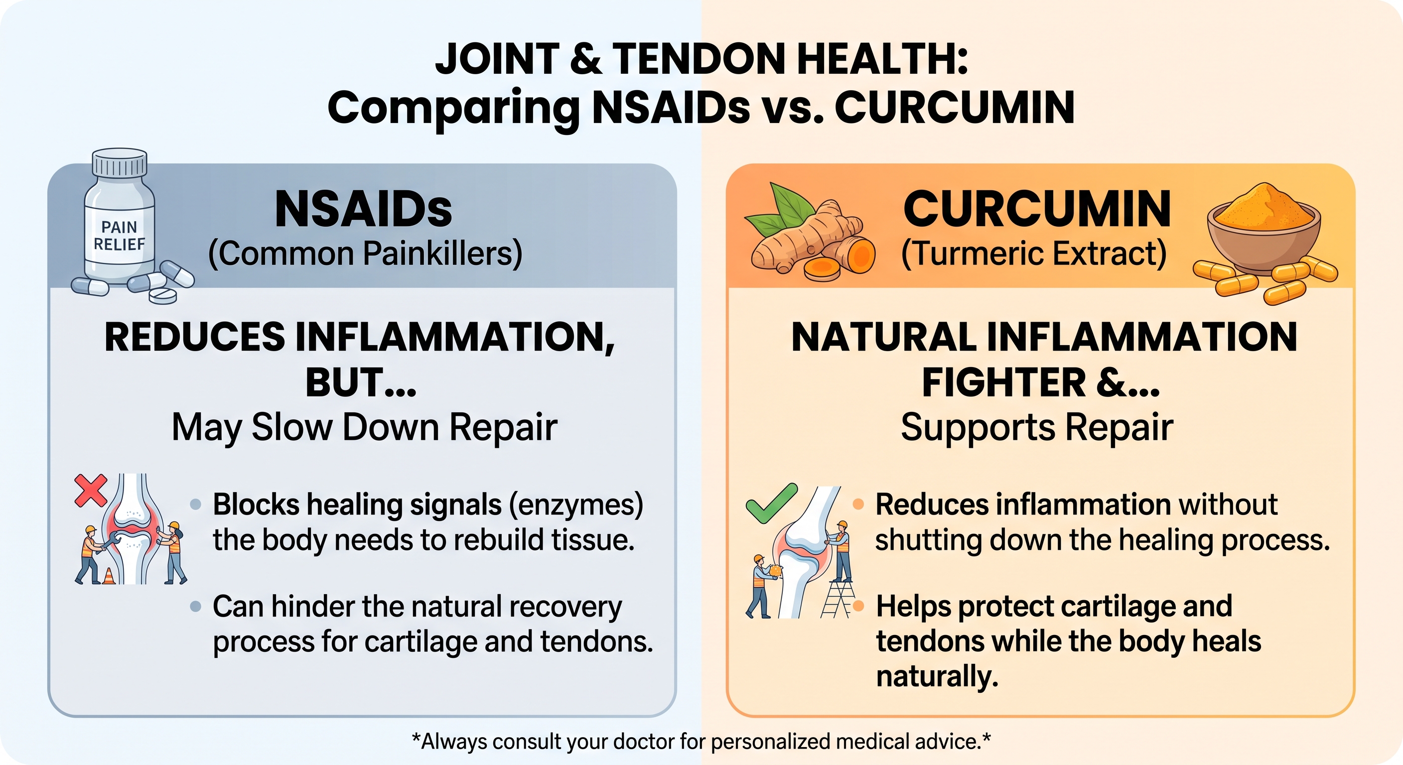
Managing soft tissue and joint injuries presents a persistent therapeutic challenge in musculoskeletal medicine. Acute inflammation triggers pain, localized swelling, and mechanical disability, which routinely drives patients toward pharmacological relief. For decades, nonsteroidal anti-inflammatory drugs (NSAIDs) such as ibuprofen, naproxen, celecoxib, and diclofenac have served as primary clinical interventions. While these synthetic agents effectively extinguish acute pain signals, expanding cellular and clinical research indicates that their biochemical mechanism interrupts the fundamental repair cascades required for long-term connective tissue healing. In contrast, curcumin - a natural polyphenolic compound derived from the rhizomes of Curcuma longa (turmeric) - exhibits a distinct, multi-targeted regulatory profile that calms hyperactive inflammation while protecting the structural components of cartilage and tendon tissue.The functional divergence between these two compounds becomes clear when considering an intuitive mechanical analogy. Tissue trauma resembles damage to a commercial building, where initial inflammation functions as the construction and demolition crew tasked with clearing rubble, stabilizing the foundation, and laying down fresh structural framing. NSAIDs function like abruptly cutting electrical power to the entire work site: the noisy machinery stops instantly and the immediate disturbance ceases, but the construction workers lose the power necessary to clean the debris and erect permanent walls. Conversely, curcumin operates as an experienced site manager: it silences unnecessary chaos and prevents site vandalism while ensuring that the skilled workers - specifically chondrocytes in cartilage and tenocytes in tendons - remain fully active, supplied, and capable of completing the restoration.
At the cellular level, NSAIDs exert their effects through the catalytic inhibition of cyclooxygenase enzymes, categorized as constitutive cyclooxygenase-1 (COX-1) and inducible cyclooxygenase-2 (COX-2). Membrane phospholipids damaged during injury release arachidonic acid, which COX enzymes convert into pro-inflammatory lipid mediators known as prostanoids, most notably prostaglandin E2 (PGE2). Nonselective NSAIDs block both isoforms, while selective coxibs specifically target COX-2. By shutting off PGE2 production, NSAIDs rapidly elevate pain thresholds and blunt localized swelling. However, because PGE2 also functions as a vital signaling cue for cellular recruitment, angiogenesis, and cellular proliferation, this total enzymatic shutdown strips local repair cells of the baseline signals required to orchestrate tissue regeneration.
Curcumin avoids this indiscriminate pathway paralysis by acting upstream on master transcriptional control switches rather than directly neutralizing isolated enzymes. Its primary therapeutic mechanism centers on the inhibition of Nuclear Factor-kappa B (NF-kB) and Activator Protein-1 (AP-1) signaling networks. Under acute stress or chronic inflammatory loads, the NF-kB protein complex is released from its cytoplasmic inhibitor, IkBa, and translocates directly into the cell nucleus, where it drives the transcription of catabolic cytokines such as interleukin-1 beta (IL-1ß), tumor necrosis factor-alpha (TNF-a), and interleukin-6 (IL-6). Curcumin blocks the phosphorylation and degradation of IkBa, effectively keeping NF-kB trapped in the cytoplasm. By preventing this nuclear entry, curcumin attenuates the expression of pro-inflammatory cytokines and downstream matrix-degrading enzymes while preserving baseline physiological functions. Furthermore, curcumin activates the nuclear factor erythroid 2-related factor 2 (Nrf2) and antioxidant response element (ARE) pathways, which upregulate endogenous cellular antioxidants to neutralize destructive reactive oxygen species (ROS) that induce cell death in injured joints.
| Pharmacological Characteristic | Nonsteroidal Anti-Inflammatory Drugs (NSAIDs) | Curcumin (Curcuma longa Polyphenol) |
| Primary Cellular Target | Direct active-site inhibition of COX-1 and/or COX-2 enzymes | Upstream inhibition of NF-kB, AP-1, and stimulation of Nrf2/ARE |
| Impact on Prostaglandin Synthesis | Profound, systemic depletion of PGE2 and related prostanoids | Modest, homeostatic modulation mediated via upstream cytokine reduction |
| Cytokine Regulation (TNF-a, IL-1ß) | Minimal direct inhibitory effect on primary cytokine gene expression | Significant downregulation of IL-1ß, TNF-a, and IL-6 secretion |
| Redox Balance and Oxidative Stress | No intrinsic reactive oxygen species scavenging capabilities | Direct free radical scavenger; stimulates endogenous antioxidant cascades |
| Organ-Level Safety Profile | Documented risks of peptic ulceration, renal stress, and cardiovascular events | Favorable tolerability profile; exhibits gastric mucosal cytoprotection |
The Mechanisms of NSAID-Induced Inhibition in Cartilage and Tendon Repair
Articular cartilage and tendons are specialized, bradytrophic connective tissues characterized by relatively low basal metabolic rates and limited endogenous vascular supplies. These physical constraints make their cellular maintenance highly vulnerable to pharmaceutical disruptions. When synthetic compounds impair cell migration, survival, or extracellular matrix secretion, the intrinsic repair capacity of these structures is substantially degraded.Cartilage maintenance depends on chondrocytes, the sole cell type residing within articular joints, which are responsible for generating and maintaining the extracellular matrix of Type II collagen and water-binding proteoglycans such as aggrecan. Because adult articular cartilage lacks a direct vascular network, major defect repair relies heavily on the recruitment and chondrogenic differentiation of subchondral mesenchymal stem cells (MSCs) through endochondral ossification. Experimental evaluations demonstrate that systemic or intra-articular NSAID exposure halts this regenerative differentiation. Both nonselective NSAIDs and selective COX-2 inhibitors interfere with chondrocyte maturation and prevent successful tissue integration following cartilage transplantation or microfracture procedures. At the cellular level, common NSAIDs - including indomethacin, ketorolac, and diclofenac - induce cell cycle arrest in chondrocytes by blocking transition from the resting G0 phase to the proliferative G1.
phase, substantially reducing viable cell numbers. Concurrently, NSAIDs downregulate Bone Morphogenetic Protein-2 (BMP-2), an essential anabolic signaling molecule that orchestrates matrix synthesis. Histological analyses in animal models reveal that NSAID administration causes a marked loss of extracellular proteoglycan content and widespread chondrocyte depletion, yielding elevated modified Mankin scores that signify advanced structural degeneration. In layman's terms, proteoglycans serve as water-absorbing structural sponges that grant cartilage its elastic shock absorption. When NSAIDs deplete these molecules, the joint surface dries out, becomes brittle, and rapidly wears down under routine mechanical friction.
Tendon healing is similarly susceptible to disruption by NSAIDs. Tendons operate as high-tensile structural cables composed of longitudinally arranged Type I collagen fibers, maintained by specialized fibroblasts termed tenocytes and tenoblasts. Healing after acute rupture or chronic tear progresses through an initial inflammatory phase, followed by a proliferative phase of cell migration, and culminates in a lengthy remodeling phase of collagen alignment. In vitro and in vivo studies establish that NSAIDs directly suppress the migration and proliferative capacity of tenocytes during the critical early healing window. Without an adequate cellular workforce migrating into the wound bed, provisional collagen scaffolding cannot be synthesized effectively.
The disruption is particularly pronounced at the enthesis, the specialized fibrocartilaginous junction where soft tendon inserts into rigid bone. Re-establishing this transition zone requires coordinated bone remodeling and chondrogenic differentiation, both of which are hindered by COX inhibition. In animal rotator cuff repair models, early administration of NSAIDs significantly delays collagen fiber organization and impairs mechanical integration at the insertion site, resulting in a measurable decline in load-to-failure strength and overall tendon toughness. Furthermore, while prolonged inflammation contributes to pathological degeneration, the transient synthesis of PGE2 during the initial hours following injury is essential for regulating localized microvascular flow and hyperemic nutrient delivery. By eliminating this early prostanoid pulse, NSAIDs starve the repair zone of oxygen and circulating factors precisely when the cellular metabolic demand is highest.
Beyond direct biochemical cytotoxicity, systemic NSAIDs introduce a physical hazard known as analgesic arthropathy. In musculoskeletal biomechanics, pain functions as a protective feedback mechanism, forcing the patient to unload an injured joint or limb to prevent structural overload. By effectively blunting the pain reflex while simultaneously undermining the cellular synthesis of proteoglycans and collagen, NSAIDs create a deceptive state of perceived recovery. Patients prematurely resume unrestricted weight-bearing and strenuous activity, placing substantial, uncompensated mechanical forces onto compromised cartilage surfaces and unhealed tendon fibers. Over time, this recurring mechanical trauma accelerates joint space narrowing, promotes microfractures in the subchondral plate, and hastens functional joint breakdown.
Chondroprotective and Tenogenic Mechanisms of Curcumin
In sharp contrast to the suppressive actions of synthetic COX inhibitors, curcumin exhibits a tissue-sparing, pro-regenerative biological profile. Rather than paralyzing cellular metabolism, curcumin modulates the microenvironment by suppressing destructive catabolic enzymes while supporting the baseline anabolic signals required for cartilage and tendon reconstruction.In articular cartilage, curcumin acts as a direct chondroprotective agent by dismantling the catabolic cascade induced by pro-inflammatory cytokines. When joint tissues are exposed to elevated IL-1ß and TNF-a, chondrocytes are provoked to synthesize matrix metalloproteinases - specifically collagenases such as MMP-1, MMP-3, and MMP-13 - along with aggrecanases like ADAMTS5. These enzymes function like enzymatic shears, systematically slicing through structural Type II collagen strands and degrading aggrecan cores. Curcumin suppresses the transcription and secretion of MMP-1, MMP-3, MMP-13, and ADAMTS5 by neutralizing NF-kB and AP-1 activation. Concurrently, it upregulates Cbp/p300 Interacting Transactivator with ED-rich tail 2 (CITED2), an essential transcriptional regulator that represses matrix metalloproteinases at the genomic level.
In addition to halting extracellular degradation, curcumin protects chondrocytes from inflammatory apoptosis. Exposure to oxidative stress and inflammatory cytokines typically triggers programmed cell death by activating the intrinsic caspase cascade. Curcumin suppresses this apoptotic pathway by reducing the cleavage of executioner caspase-3, downregulating the pro-apoptotic factor Bax, and elevating anti-apoptotic Bcl-2 expression. Microscopic and histological analyses of osteoarthritic cartilage treated with curcumin consistently show robust Safranin O staining, intact surface regularity, preserved chondrocyte density, and significantly reduced Osteoarthritis Research Society International (OARSI) degradation scores.
Within tendon biology, curcumin promotes active tissue regeneration (tenogenesis) rather than inert scar formation. In animal models of tendon rupture and surgical repair, curcumin guides tendon-derived stem and progenitor cells (TSPCs) toward mature tenocyte differentiation. This lineage-specific differentiation is orchestrated primarily through activation of the phosphatidylinositol 3-kinase/protein kinase B (PI3K/Akt) and Wnt/ß-catenin signaling cascades. As stem cells commit to the tenogenic pathway, they significantly upregulate crucial structural and phenotypic markers, including epithelial cell adhesion molecule (EpCAM), tenomodulin, and the master tendon transcription factor Scleraxis.
Through this guided differentiation, curcumin supports the preferential synthesis and organized deposition of high-tensile Type I collagen fibers, the primary architectural building blocks of healthy tendons. Injured tendons typically fill with disorganized, loose Type III collagen, which forms a structurally inferior, compliant scar. Curcumin supplementation promotes the progressive maturation from primitive Type III collagen to densely packed, parallel Type I collagen fibrils, restoring native tensile strength and mechanical breaking force.
A major clinical challenge following tendon surgery is the development of peritendinous adhesions, in which excessive fibrotic scar tissue binds the healing tendon to its synovial sheath, restricting range of motion and joint gliding. Postoperative tendon repair involves a balance between extrinsic healing (in which exterior fibroblasts infiltrate the defect and deposit messy scar tissue) and intrinsic healing (in which tenocytes within the tendon substance reconstruct the matrix). Curcumin suppresses excessive peritendinous inflammation and extrinsic fibrosis while accelerating intrinsic tenocyte repair. Biomechanical testing confirms that local or systemic curcumin administration yields lower adhesion scores, increased gliding distances, and superior functional mobility without sacrificing structural load-bearing capacity.
Comparative Clinical Efficacy and Systemic Safety Profiles
Translating cellular findings into clinical practice requires weighing therapeutic outcomes against systemic safety profiles. While synthetic NSAIDs deliver potent, rapid analgesia during acute musculoskeletal pain events, their chronic administration is constrained by systemic toxicities across multiple organ systems.In randomized, active-controlled clinical trials of knee osteoarthritis, optimized curcumin preparations have demonstrated clinical pain reduction and functional recovery comparable to standard therapeutic doses of NSAIDs. In a randomized, open-label parallel-arm study evaluating 139 patients with symptomatic knee osteoarthritis, subjects received either 500 mg of a bio-enhanced curcumin extract three times daily or 50 mg of diclofenac sodium twice daily for 28 consecutive days. Patients treated with curcumin demonstrated comparable improvements in pain intensity on the Visual Analogue Scale (VAS) and functional scores on the Knee Injury and Osteoarthritis Outcome Score (KOOS) at days 14 and 28, showing no statistically significant difference in therapeutic efficacy compared to diclofenac.
However, the systemic tolerability profiles between the treatments diverged markedly. Overall adverse events occurred in only 13% of the curcumin group compared to 38% of the diclofenac cohort. In the diclofenac arm, 28% of patients developed dyspeptic symptoms severe enough to require concurrent treatment with H2-receptor antagonists or proton pump inhibitors to prevent mucosal ulceration, whereas no patients in the curcumin cohort required gastroprotective intervention. Furthermore, the curcumin cohort experienced a significant reduction in flatulence and digestive discomfort, demonstrating beneficial gastric and intestinal cytoprotective effects.
These safety observations are consistent across broader systematic reviews and meta-analyses. Nonsteroidal anti-inflammatory agents inherently compromise gastrointestinal integrity by systematically depleting cytoprotective prostaglandins, leaving the gastric epithelium vulnerable to acid erosion and hemorrhage. In addition, systemic inhibition of renal and vascular COX enzymes by NSAIDs reduces renal perfusion and disrupts the balance between prostacyclin and thromboxane, elevating the risk of fluid retention, hypertension, and adverse cardiovascular thrombotic events. Curcumin does not suppress these physiological prostanoid pathways, making it free from ulcerogenic, nephrotoxic, and cardiotoxic properties at therapeutic dosages.
| Clinical Parameter | Conventional NSAIDs (e.g., Diclofenac, Ibuprofen) | Formulated Curcumin (Curcuma longa) |
| Onset and Depth of Analgesia | Rapid onset (1 to 2 hours); potent, broad suppression of acute pain | Progressive onset (several days); steady, moderate-to-high pain relief |
| Functional Joint Scores (KOOS / WOMAC) | Significant, established improvements in mobility and stiffness scores | Statistically comparable improvements in functional and quality-of-life scores |
| Gastrointestinal Integrity | Elevated incidence of dyspepsia, gastric erosions, and peptic ulcer bleeding | Gastroprotective; exhibits anti-ulcer actions and improves digestive tolerance |
| Cardiovascular and Renal Strain | Documented risks of fluid retention, renal dysfunction, and thrombotic events | Favorable safety profile; provides systemic antioxidant and vascular benefits |
| Cartilage Matrix Dynamics | Associated with proteoglycan depletion, cell arrest, and matrix breakdown | Chondroprotective; inhibits MMPs/ADAMTS5 while preserving proteoglycans |
| Tendon Remodeling and Strength | Suppresses tenocyte migration and compromises enthesis breaking strength | Enhances tenogenesis, organizes Type I collagen, and limits adhesion formation |
A longstanding limitation of standard curcumin supplementation in clinical settings has been its low oral bioavailability. Native curcuminoids are highly lipophilic, poorly soluble in aqueous gastric fluids, and subject to rapid hepatic and intestinal phase II metabolism into inactive glucuronides and sulfates, leading to swift biliary and fecal excretion. Consequently, raw culinary turmeric powders struggle to achieve the therapeutic systemic circulating concentrations required to reach poorly vascularized joint and tendon compartments. Modern pharmacognosy has addressed this pharmacodynamic hurdle through advanced delivery formulations. Modern strategies - such as co-administration with the natural alkaloid piperine (which inhibits hepatic glucuronidation), micellar dispersions, phytosomal phospholipid complexes, and bio-enhanced submicron dispersions - elevate systemic blood bioavailability by ten- to thirty-fold compared to unformulated extracts. These modern delivery platforms ensure that therapeutic concentrations reach synovial fluid, subchondral bone, and fibrous tendon sheaths without requiring excessive oral dosing.
Conclusions and Translational Clinical Implications
The biological and clinical evidence reveals distinct physiological paths for NSAIDs and curcumin in orthopedic recovery. While NSAIDs remain powerful tools for the brief alleviation of acute, unmanageable pain, their ongoing administration during active tissue healing presents substantial biological compromises. Tendons and cartilage require a controlled, transient inflammatory cascade to signal cell recruitment, stimulate stem cell differentiation, and direct extracellular matrix synthesis. By completely shutting down cyclooxygenase enzymes and depleting local prostaglandins, NSAIDs disrupt this regenerative cascade. The resulting cellular consequences - including chondrocyte cell cycle arrest, proteoglycan loss, suppression of tenocyte migration, and weakened collagen tensile strength at the tendon-to-bone interface - demonstrate that pain relief from NSAIDs frequently comes at the cost of the structural integrity of healing connective tissues.In contrast, curcumin provides a tissue-preserving alternative that decouples pain and inflammation control from cellular suppression. Operating upstream at the level of NF-kB and AP-1 transcriptional activation, curcumin attenuates the expression of pro-inflammatory cytokines while leaving the physiological baseline of cellular metabolism intact. In cartilage, it actively represses the matrix-degrading enzymes MMP-1, MMP-3, MMP-13, and ADAMTS5, maintains water-binding proteoglycan content, and protects chondrocytes from inflammatory apoptosis. In damaged tendons, curcumin stimulates tenogenic differentiation of local stem cells via PI3K/Akt signaling, supports the proper maturation of dense Type I collagen fibers, and prevents restrictive peritendinous scar adhesions, preserving both joint mobility and mechanical breaking strength.
From a translational perspective, these findings indicate that clinical protocols should reconsider relying on continuous NSAIDs as the default intervention for connective tissue injuries, post-surgical recovery, and chronic degenerative conditions. Where synthetic NSAIDs are deemed necessary, their use should be confined to short-term acute flare-ups to avoid interrupting early tissue remodeling. For long-term joint preservation, ongoing tendinopathy rehabilitation, and chronic osteoarthritis management, optimized bio-enhanced curcumin formulations deliver pain relief and functional restoration comparable to conventional pharmaceuticals, all while preserving the biological processes required for lasting musculoskeletal repair.
References:
NSAID therapy effects on healing of bone, tendon, and the enthesis
The Detrimental Effects of Systemic Ibuprofen Delivery on Tendon
Conservative management of symptomatic knee osteoarthritis - PMC
Positives and negatives of nonsteroidal anti-inflammatory drugs in
(PDF) Conservative management of symptomatic knee osteoarthritis
Non-steroidal anti-inflammatory drugs influence cartilage healing
Is Curcumine Useful in the Treatment and Prevention of ... - PMC - NIH
Efficacy and mechanisms of curcumin in the treatment of osteoarthritis
Biological actions of curcumin on articular chondrocytes - PubMed
Curcumin slows osteoarthritis progression and relieves ... - PMC - NIH
Do Nonsteroidal Anti-Inflammatory Drugs Have a Deleterious Effect
Anti-inflammatory management for tendon injuries - friends or foes?
The mechanistic role of curcumin on matrix metalloproteinases in
Therapeutic effects of turmeric or curcumin extract on pain and
Curcumin inhibits chondrocyte apoptosis and inflammation in ... - PMC
Analysis of the mechanism of curcumin against osteoarthritis using
Synergistic enhancement of tendon-to-bone healing via anti ... - PMC
A Multicenter, Randomized, Double-Blind, Placebo-Controlled Trial
Safety and efficacy of curcumin versus diclofenac in knee osteoarthritis
A randomized, pilot study to assess the efficacy and safety ... - PubMed
Effectiveness of curcuminoids in the treatment of knee osteoarthritis
THE EFFECTS OF TRADITIONAL STRENGTHENING EXERCISES
Putting Some Muscle into Osteoarthritis | Annals of Internal Medicine
Do Nonsteroidal Anti-Inflammatory Drugs Have a Deleterious Effect
Calebin A, a Compound of Turmeric, Down-Regulates Inflammation
The Effect of Non-Steroidal Anti-Inflammatory Drugs on Tendon-to
Chiropractor in Toronto, ON, Canada :: Head and Neck Pain
Investigation of the association of long-term NSAID use with ... - PMC
Curcumin Improves Functional Recovery of Ruptured Tendon by
Chemically modified curcumin (CMC2.24) alleviates osteoarthritis
Curcumin improves age-related and surgically induced osteoarthritis
(PDF) Curcumin Improves Functional Recovery of Ruptured Tendon
Investigation of the effects of umbilical cord-derived mesenchymal
Effect of curcumin-loaded polycaprolactone scaffold on Achilles
Controlled release of curcumin from curcumin-loaded nanomicelles
Efficacy and safety of combination of curcuminoid complex ... - PMC
The efficacy of Curcuma Longa L. extract as an adjuvant therapy in