|
Search Term: " Dino "
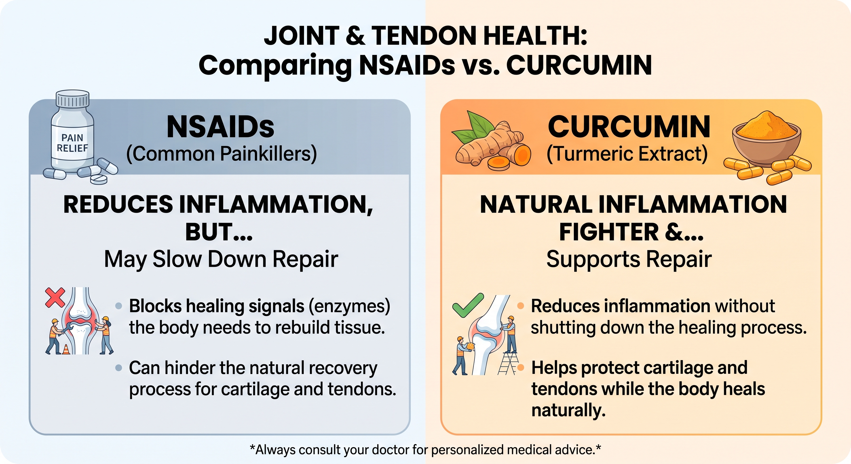
NSAIDs vs. Curcumin: Which One Relieves Joint Pain Without Stopping Healing?   
Date:
September 14, 2026 11:22 AM
Comparative Analysis of NSAIDs versus Curcumin in Musculoskeletal Tissue RepairPrimary Biological Mechanisms and Pharmacological TargetsManaging soft tissue and joint injuries presents a persistent therapeutic challenge in musculoskeletal medicine. Acute inflammation triggers pain, localized swelling, and mechanical disability, which routinely drives patients toward pharmacological relief. For decades, nonsteroidal anti-inflammatory drugs (NSAIDs) such as ibuprofen, naproxen, celecoxib, and diclofenac have served as primary clinical interventions. While these synthetic agents effectively extinguish acute pain signals, expanding cellular and clinical research indicates that their biochemical mechanism interrupts the fundamental repair cascades required for long-term connective tissue healing. In contrast, curcumin - a natural polyphenolic compound derived from the rhizomes of Curcuma longa (turmeric) - exhibits a distinct, multi-targeted regulatory profile that calms hyperactive inflammation while protecting the structural components of cartilage and tendon tissue.The functional divergence between these two compounds becomes clear when considering an intuitive mechanical analogy. Tissue trauma resembles damage to a commercial building, where initial inflammation functions as the construction and demolition crew tasked with clearing rubble, stabilizing the foundation, and laying down fresh structural framing. NSAIDs function like abruptly cutting electrical power to the entire work site: the noisy machinery stops instantly and the immediate disturbance ceases, but the construction workers lose the power necessary to clean the debris and erect permanent walls. Conversely, curcumin operates as an experienced site manager: it silences unnecessary chaos and prevents site vandalism while ensuring that the skilled workers - specifically chondrocytes in cartilage and tenocytes in tendons - remain fully active, supplied, and capable of completing the restoration. At the cellular level, NSAIDs exert their effects through the catalytic inhibition of cyclooxygenase enzymes, categorized as constitutive cyclooxygenase-1 (COX-1) and inducible cyclooxygenase-2 (COX-2). Membrane phospholipids damaged during injury release arachidonic acid, which COX enzymes convert into pro-inflammatory lipid mediators known as prostanoids, most notably prostaglandin E2 (PGE2). Nonselective NSAIDs block both isoforms, while selective coxibs specifically target COX-2. By shutting off PGE2 production, NSAIDs rapidly elevate pain thresholds and blunt localized swelling. However, because PGE2 also functions as a vital signaling cue for cellular recruitment, angiogenesis, and cellular proliferation, this total enzymatic shutdown strips local repair cells of the baseline signals required to orchestrate tissue regeneration. Curcumin avoids this indiscriminate pathway paralysis by acting upstream on master transcriptional control switches rather than directly neutralizing isolated enzymes. Its primary therapeutic mechanism centers on the inhibition of Nuclear Factor-kappa B (NF-kB) and Activator Protein-1 (AP-1) signaling networks. Under acute stress or chronic inflammatory loads, the NF-kB protein complex is released from its cytoplasmic inhibitor, IkBa, and translocates directly into the cell nucleus, where it drives the transcription of catabolic cytokines such as interleukin-1 beta (IL-1ß), tumor necrosis factor-alpha (TNF-a), and interleukin-6 (IL-6). Curcumin blocks the phosphorylation and degradation of IkBa, effectively keeping NF-kB trapped in the cytoplasm. By preventing this nuclear entry, curcumin attenuates the expression of pro-inflammatory cytokines and downstream matrix-degrading enzymes while preserving baseline physiological functions. Furthermore, curcumin activates the nuclear factor erythroid 2-related factor 2 (Nrf2) and antioxidant response element (ARE) pathways, which upregulate endogenous cellular antioxidants to neutralize destructive reactive oxygen species (ROS) that induce cell death in injured joints.
The Mechanisms of NSAID-Induced Inhibition in Cartilage and Tendon RepairArticular cartilage and tendons are specialized, bradytrophic connective tissues characterized by relatively low basal metabolic rates and limited endogenous vascular supplies. These physical constraints make their cellular maintenance highly vulnerable to pharmaceutical disruptions. When synthetic compounds impair cell migration, survival, or extracellular matrix secretion, the intrinsic repair capacity of these structures is substantially degraded.Cartilage maintenance depends on chondrocytes, the sole cell type residing within articular joints, which are responsible for generating and maintaining the extracellular matrix of Type II collagen and water-binding proteoglycans such as aggrecan. Because adult articular cartilage lacks a direct vascular network, major defect repair relies heavily on the recruitment and chondrogenic differentiation of subchondral mesenchymal stem cells (MSCs) through endochondral ossification. Experimental evaluations demonstrate that systemic or intra-articular NSAID exposure halts this regenerative differentiation. Both nonselective NSAIDs and selective COX-2 inhibitors interfere with chondrocyte maturation and prevent successful tissue integration following cartilage transplantation or microfracture procedures. At the cellular level, common NSAIDs - including indomethacin, ketorolac, and diclofenac - induce cell cycle arrest in chondrocytes by blocking transition from the resting G0 phase to the proliferative G1. phase, substantially reducing viable cell numbers. Concurrently, NSAIDs downregulate Bone Morphogenetic Protein-2 (BMP-2), an essential anabolic signaling molecule that orchestrates matrix synthesis. Histological analyses in animal models reveal that NSAID administration causes a marked loss of extracellular proteoglycan content and widespread chondrocyte depletion, yielding elevated modified Mankin scores that signify advanced structural degeneration. In layman's terms, proteoglycans serve as water-absorbing structural sponges that grant cartilage its elastic shock absorption. When NSAIDs deplete these molecules, the joint surface dries out, becomes brittle, and rapidly wears down under routine mechanical friction. Tendon healing is similarly susceptible to disruption by NSAIDs. Tendons operate as high-tensile structural cables composed of longitudinally arranged Type I collagen fibers, maintained by specialized fibroblasts termed tenocytes and tenoblasts. Healing after acute rupture or chronic tear progresses through an initial inflammatory phase, followed by a proliferative phase of cell migration, and culminates in a lengthy remodeling phase of collagen alignment. In vitro and in vivo studies establish that NSAIDs directly suppress the migration and proliferative capacity of tenocytes during the critical early healing window. Without an adequate cellular workforce migrating into the wound bed, provisional collagen scaffolding cannot be synthesized effectively. The disruption is particularly pronounced at the enthesis, the specialized fibrocartilaginous junction where soft tendon inserts into rigid bone. Re-establishing this transition zone requires coordinated bone remodeling and chondrogenic differentiation, both of which are hindered by COX inhibition. In animal rotator cuff repair models, early administration of NSAIDs significantly delays collagen fiber organization and impairs mechanical integration at the insertion site, resulting in a measurable decline in load-to-failure strength and overall tendon toughness. Furthermore, while prolonged inflammation contributes to pathological degeneration, the transient synthesis of PGE2 during the initial hours following injury is essential for regulating localized microvascular flow and hyperemic nutrient delivery. By eliminating this early prostanoid pulse, NSAIDs starve the repair zone of oxygen and circulating factors precisely when the cellular metabolic demand is highest. Beyond direct biochemical cytotoxicity, systemic NSAIDs introduce a physical hazard known as analgesic arthropathy. In musculoskeletal biomechanics, pain functions as a protective feedback mechanism, forcing the patient to unload an injured joint or limb to prevent structural overload. By effectively blunting the pain reflex while simultaneously undermining the cellular synthesis of proteoglycans and collagen, NSAIDs create a deceptive state of perceived recovery. Patients prematurely resume unrestricted weight-bearing and strenuous activity, placing substantial, uncompensated mechanical forces onto compromised cartilage surfaces and unhealed tendon fibers. Over time, this recurring mechanical trauma accelerates joint space narrowing, promotes microfractures in the subchondral plate, and hastens functional joint breakdown. Chondroprotective and Tenogenic Mechanisms of CurcuminIn sharp contrast to the suppressive actions of synthetic COX inhibitors, curcumin exhibits a tissue-sparing, pro-regenerative biological profile. Rather than paralyzing cellular metabolism, curcumin modulates the microenvironment by suppressing destructive catabolic enzymes while supporting the baseline anabolic signals required for cartilage and tendon reconstruction.In articular cartilage, curcumin acts as a direct chondroprotective agent by dismantling the catabolic cascade induced by pro-inflammatory cytokines. When joint tissues are exposed to elevated IL-1ß and TNF-a, chondrocytes are provoked to synthesize matrix metalloproteinases - specifically collagenases such as MMP-1, MMP-3, and MMP-13 - along with aggrecanases like ADAMTS5. These enzymes function like enzymatic shears, systematically slicing through structural Type II collagen strands and degrading aggrecan cores. Curcumin suppresses the transcription and secretion of MMP-1, MMP-3, MMP-13, and ADAMTS5 by neutralizing NF-kB and AP-1 activation. Concurrently, it upregulates Cbp/p300 Interacting Transactivator with ED-rich tail 2 (CITED2), an essential transcriptional regulator that represses matrix metalloproteinases at the genomic level. In addition to halting extracellular degradation, curcumin protects chondrocytes from inflammatory apoptosis. Exposure to oxidative stress and inflammatory cytokines typically triggers programmed cell death by activating the intrinsic caspase cascade. Curcumin suppresses this apoptotic pathway by reducing the cleavage of executioner caspase-3, downregulating the pro-apoptotic factor Bax, and elevating anti-apoptotic Bcl-2 expression. Microscopic and histological analyses of osteoarthritic cartilage treated with curcumin consistently show robust Safranin O staining, intact surface regularity, preserved chondrocyte density, and significantly reduced Osteoarthritis Research Society International (OARSI) degradation scores. Within tendon biology, curcumin promotes active tissue regeneration (tenogenesis) rather than inert scar formation. In animal models of tendon rupture and surgical repair, curcumin guides tendon-derived stem and progenitor cells (TSPCs) toward mature tenocyte differentiation. This lineage-specific differentiation is orchestrated primarily through activation of the phosphatidylinositol 3-kinase/protein kinase B (PI3K/Akt) and Wnt/ß-catenin signaling cascades. As stem cells commit to the tenogenic pathway, they significantly upregulate crucial structural and phenotypic markers, including epithelial cell adhesion molecule (EpCAM), tenomodulin, and the master tendon transcription factor Scleraxis. Through this guided differentiation, curcumin supports the preferential synthesis and organized deposition of high-tensile Type I collagen fibers, the primary architectural building blocks of healthy tendons. Injured tendons typically fill with disorganized, loose Type III collagen, which forms a structurally inferior, compliant scar. Curcumin supplementation promotes the progressive maturation from primitive Type III collagen to densely packed, parallel Type I collagen fibrils, restoring native tensile strength and mechanical breaking force. A major clinical challenge following tendon surgery is the development of peritenDinous adhesions, in which excessive fibrotic scar tissue binds the healing tendon to its synovial sheath, restricting range of motion and joint gliding. Postoperative tendon repair involves a balance between extrinsic healing (in which exterior fibroblasts infiltrate the defect and deposit messy scar tissue) and intrinsic healing (in which tenocytes within the tendon substance reconstruct the matrix). Curcumin suppresses excessive peritenDinous inflammation and extrinsic fibrosis while accelerating intrinsic tenocyte repair. Biomechanical testing confirms that local or systemic curcumin administration yields lower adhesion scores, increased gliding distances, and superior functional mobility without sacrificing structural load-bearing capacity. Comparative Clinical Efficacy and Systemic Safety ProfilesTranslating cellular findings into clinical practice requires weighing therapeutic outcomes against systemic safety profiles. While synthetic NSAIDs deliver potent, rapid analgesia during acute musculoskeletal pain events, their chronic administration is constrained by systemic toxicities across multiple organ systems.In randomized, active-controlled clinical trials of knee osteoarthritis, optimized curcumin preparations have demonstrated clinical pain reduction and functional recovery comparable to standard therapeutic doses of NSAIDs. In a randomized, open-label parallel-arm study evaluating 139 patients with symptomatic knee osteoarthritis, subjects received either 500 mg of a bio-enhanced curcumin extract three times daily or 50 mg of diclofenac sodium twice daily for 28 consecutive days. Patients treated with curcumin demonstrated comparable improvements in pain intensity on the Visual Analogue Scale (VAS) and functional scores on the Knee Injury and Osteoarthritis Outcome Score (KOOS) at days 14 and 28, showing no statistically significant difference in therapeutic efficacy compared to diclofenac. However, the systemic tolerability profiles between the treatments diverged markedly. Overall adverse events occurred in only 13% of the curcumin group compared to 38% of the diclofenac cohort. In the diclofenac arm, 28% of patients developed dyspeptic symptoms severe enough to require concurrent treatment with H2-receptor antagonists or proton pump inhibitors to prevent mucosal ulceration, whereas no patients in the curcumin cohort required gastroprotective intervention. Furthermore, the curcumin cohort experienced a significant reduction in flatulence and digestive discomfort, demonstrating beneficial gastric and intestinal cytoprotective effects. These safety observations are consistent across broader systematic reviews and meta-analyses. Nonsteroidal anti-inflammatory agents inherently compromise gastrointestinal integrity by systematically depleting cytoprotective prostaglandins, leaving the gastric epithelium vulnerable to acid erosion and hemorrhage. In addition, systemic inhibition of renal and vascular COX enzymes by NSAIDs reduces renal perfusion and disrupts the balance between prostacyclin and thromboxane, elevating the risk of fluid retention, hypertension, and adverse cardiovascular thrombotic events. Curcumin does not suppress these physiological prostanoid pathways, making it free from ulcerogenic, nephrotoxic, and cardiotoxic properties at therapeutic dosages.
A longstanding limitation of standard curcumin supplementation in clinical settings has been its low oral bioavailability. Native curcuminoids are highly lipophilic, poorly soluble in aqueous gastric fluids, and subject to rapid hepatic and intestinal phase II metabolism into inactive glucuronides and sulfates, leading to swift biliary and fecal excretion. Consequently, raw culinary turmeric powders struggle to achieve the therapeutic systemic circulating concentrations required to reach poorly vascularized joint and tendon compartments. Modern pharmacognosy has addressed this pharmacodynamic hurdle through advanced delivery formulations. Modern strategies - such as co-administration with the natural alkaloid piperine (which inhibits hepatic glucuronidation), micellar dispersions, phytosomal phospholipid complexes, and bio-enhanced submicron dispersions - elevate systemic blood bioavailability by ten- to thirty-fold compared to unformulated extracts. These modern delivery platforms ensure that therapeutic concentrations reach synovial fluid, subchondral bone, and fibrous tendon sheaths without requiring excessive oral dosing. Conclusions and Translational Clinical ImplicationsThe biological and clinical evidence reveals distinct physiological paths for NSAIDs and curcumin in orthopedic recovery. While NSAIDs remain powerful tools for the brief alleviation of acute, unmanageable pain, their ongoing administration during active tissue healing presents substantial biological compromises. Tendons and cartilage require a controlled, transient inflammatory cascade to signal cell recruitment, stimulate stem cell differentiation, and direct extracellular matrix synthesis. By completely shutting down cyclooxygenase enzymes and depleting local prostaglandins, NSAIDs disrupt this regenerative cascade. The resulting cellular consequences - including chondrocyte cell cycle arrest, proteoglycan loss, suppression of tenocyte migration, and weakened collagen tensile strength at the tendon-to-bone interface - demonstrate that pain relief from NSAIDs frequently comes at the cost of the structural integrity of healing connective tissues.In contrast, curcumin provides a tissue-preserving alternative that decouples pain and inflammation control from cellular suppression. Operating upstream at the level of NF-kB and AP-1 transcriptional activation, curcumin attenuates the expression of pro-inflammatory cytokines while leaving the physiological baseline of cellular metabolism intact. In cartilage, it actively represses the matrix-degrading enzymes MMP-1, MMP-3, MMP-13, and ADAMTS5, maintains water-binding proteoglycan content, and protects chondrocytes from inflammatory apoptosis. In damaged tendons, curcumin stimulates tenogenic differentiation of local stem cells via PI3K/Akt signaling, supports the proper maturation of dense Type I collagen fibers, and prevents restrictive peritenDinous scar adhesions, preserving both joint mobility and mechanical breaking strength. From a translational perspective, these findings indicate that clinical protocols should reconsider relying on continuous NSAIDs as the default intervention for connective tissue injuries, post-surgical recovery, and chronic degenerative conditions. Where synthetic NSAIDs are deemed necessary, their use should be confined to short-term acute flare-ups to avoid interrupting early tissue remodeling. For long-term joint preservation, ongoing tenDinopathy rehabilitation, and chronic osteoarthritis management, optimized bio-enhanced curcumin formulations deliver pain relief and functional restoration comparable to conventional pharmaceuticals, all while preserving the biological processes required for lasting musculoskeletal repair. References: NSAID therapy effects on healing of bone, tendon, and the enthesis The Detrimental Effects of Systemic Ibuprofen Delivery on Tendon Conservative management of symptomatic knee osteoarthritis - PMC Positives and negatives of nonsteroidal anti-inflammatory drugs in (PDF) Conservative management of symptomatic knee osteoarthritis Non-steroidal anti-inflammatory drugs influence cartilage healing Is Curcumine Useful in the Treatment and Prevention of ... - PMC - NIH Efficacy and mechanisms of curcumin in the treatment of osteoarthritis Biological actions of curcumin on articular chondrocytes - PubMed Curcumin slows osteoarthritis progression and relieves ... - PMC - NIH Do Nonsteroidal Anti-Inflammatory Drugs Have a Deleterious Effect Anti-inflammatory management for tendon injuries - friends or foes? The mechanistic role of curcumin on matrix metalloproteinases in Therapeutic effects of turmeric or curcumin extract on pain and Curcumin inhibits chondrocyte apoptosis and inflammation in ... - PMC Analysis of the mechanism of curcumin against osteoarthritis using Synergistic enhancement of tendon-to-bone healing via anti ... - PMC A Multicenter, Randomized, Double-Blind, Placebo-Controlled Trial Safety and efficacy of curcumin versus diclofenac in knee osteoarthritis A randomized, pilot study to assess the efficacy and safety ... - PubMed Effectiveness of curcuminoids in the treatment of knee osteoarthritis THE EFFECTS OF TRADITIONAL STRENGTHENING EXERCISES Putting Some Muscle into Osteoarthritis | Annals of Internal Medicine Do Nonsteroidal Anti-Inflammatory Drugs Have a Deleterious Effect Calebin A, a Compound of Turmeric, Down-Regulates Inflammation The Effect of Non-Steroidal Anti-Inflammatory Drugs on Tendon-to Chiropractor in Toronto, ON, Canada :: Head and Neck Pain Investigation of the association of long-term NSAID use with ... - PMC Curcumin Improves Functional Recovery of Ruptured Tendon by Chemically modified curcumin (CMC2.24) alleviates osteoarthritis Curcumin improves age-related and surgically induced osteoarthritis (PDF) Curcumin Improves Functional Recovery of Ruptured Tendon Investigation of the effects of umbilical cord-derived mesenchymal Effect of curcumin-loaded polycaprolactone scaffold on Achilles Controlled release of curcumin from curcumin-loaded nanomicelles Efficacy and safety of combination of curcuminoid complex ... - PMC The efficacy of Curcuma Longa L. extract as an adjuvant therapy in
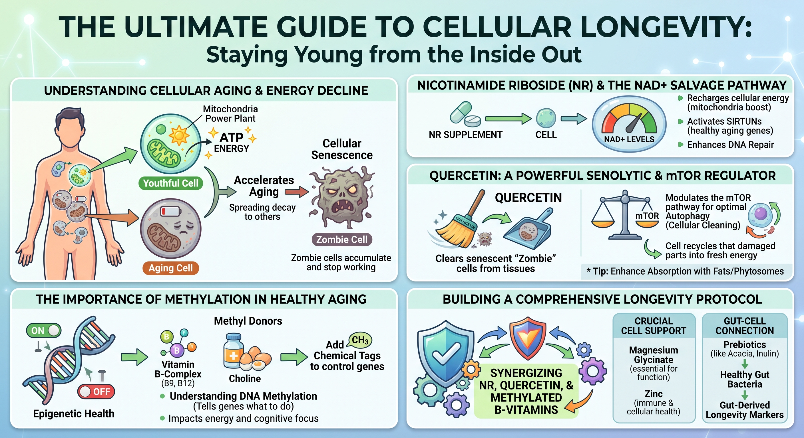
(https://vitanetonline.com:443/forums/Index.cfm?CFApp=1&Message_ID=6653) The Ultimate Guide to Cellular Longevity: NAD+, Methylation, and Senolytics 
Date:
September 10, 2026 10:57 AM
Introduction: Understanding Cellular Aging and Energy DeclineBiological aging represents a progressive decline in cellular maintenance, structural repair, and energy generation. Over decades, tissues experience an attrition of functional reserves, compromised stress resilience, and persistent low-grade systemic inflammation. At the cellular scale, biological degeneration is driven by a failure to generate bioenergetic fuel, repair genetic code, and clear metabolic waste.Cellular aging is characterized by interconnected biological disruptions known as the hallmarks of aging. These encompass genomic instability, epigenetic alterations, mitochondrial decay, loss of proteostasis, and cellular senescence. Rather than operating as isolated occurrences, these phenomena establish a self-reinforcing degenerative cycle: declining cellular power generation impairs enzymatic genetic repair, promoting the accumulation of damaged cells that enter irreversible growth arrest and poison surrounding healthy tissues. Mitigating cellular aging requires examining how microscopic bioenergetic pathways deteriorate and evaluating how targeted nutritional and biochemical interventions can restore cellular homeostasis. The Role of Mitochondria and ATP ProductionEvery biological function - from muscular contraction to continuous DNA replication - depends on adenosine triphosphate (ATP), the primary biochemical energy currency of living systems. Cells produce the vast majority of this energy within mitochondria through oxidative phosphorylation. Within these specialized organelles, metabolic intermediates derived from dietary carbohydrates and lipids donate high-energy electrons to the electron transport chain. The flow of these electrons across protein complexes establishes an electrochemical proton gradient across the inner mitochondrial membrane, driving ATP synthase to manufacture ATP.A youthful cell functions like an efficient municipal power grid, dynamically matching energetic demands with immediate ATP output. However, as biological aging progresses, mitochondrial efficiency declines. The electron transport chain becomes structurally leaky, inadvertently shedding electrons that react with ambient molecular oxygen to produce reactive oxygen species (ROS). While regulated levels of ROS participate in vital intracellular signaling, chronic excess induces widespread oxidative stress. Mitochondria are exceptionally vulnerable to this oxidative burden because they carry their own circular genetic material, known as mitochondrial DNA (mtDNA). Unlike nuclear DNA, mtDNA lacks the protective shielding of histone proteins and possesses rudimentary repair systems. As a result, mtDNA sustains cumulative oxidative damage, encoding increasingly defective electron transport chain proteins. This dynamic generates a bioenergetic deficit: degraded mitochondria synthesize progressively less ATP while emitting greater volumes of damaging free radicals. Deprived of optimal ATP reserves, cells lack the energy necessary to drive vital enzymatic repair cascades, accelerating structural degeneration and functional exhaustion. How Cellular Senescence Accelerates the Aging ProcessWhen healthy cells confront critical physiological damage - such as severe telomere attrition, persistent DNA double-strand breaks, or oxidative stress - they activate protective cell cycle arrest pathways governed primarily by the p53/p21^CIP1 and p16^INK4a/Rb molecular checkpoints. This defensive shutdown, termed cellular senescence, permanently prevents the replication of potentially premalignant or mutated cells.Senescent cells, colloquially known as "zombie cells," enter a state of permanent growth arrest while actively resisting programmed cell death (apoptosis). Over time, these cells accumulate within adipose depots, skeletal muscle, the vascular endothelium, and major organs, largely because immune surveillance and clearance pathways simultaneously lose functional efficiency. The systemic danger of senescent cells stems from their secretome. Rather than remaining biologically inert, senescent cells develop a hyperactive secretory state termed the Senescence-Associated Secretory Phenotype (SASP). The SASP is a destructive mixture of pro-inflammatory cytokines, chemokines, extracellular matrix-degrading matrix metalloproteinases (MMPs), and reactive oxygen species. Through this toxic secretome, even a small burden of senescent cells can impair whole-tissue architecture. SASP factors degrade surrounding structural proteins, induce insulin resistance in neighboring metabolic cells, and biochemically force adjacent healthy cells into secondary senescence. This persistent paracrine signaling fuels chronic, sterile, low-grade systemic inflammation, termed "inflammaging," which accelerates systemic tissue degeneration and elevates susceptibility to degenerative age-related pathologies. Nicotinamide Riboside (NR) and the NAD+ Salvage PathwayThe Biochemistry of NAD+ Depletion Over TimeNicotinamide adenine dinucleotide (NAD+) is an indispensable coenzyme present in every living cell. NAD+ fulfills a dual biological mandate: it serves as a central redox cofactor that shuttles electrons between cellular metabolic reactions, and it functions as an obligatory consumable substrate for regulatory enzymes that preserve cellular viability. In its redox capacity, NAD+ accepts electrons to form NADH during glycolysis, the tricarboxylic acid (TCA) cycle, and fatty acid beta-oxidation, subsequently donating those electrons to Complex I of the respiratory chain to power ATP synthesis.
The primary enzymatic driver of age-related NAD+ destruction is CD38, a membrane-bound glycohydrolase expressed on immune cells that is upregulated in response to chronic SASP exposure. Concurrently, lifelong genotoxic damage causes persistent activation of Poly(ADP-ribose) polymerase 1 (PARP-1), an enzyme that cleaves the glycosidic bonds of NAD+ to assemble branched poly(ADP-ribose) chains at DNA lesion sites. Because PARP-1 consumes NAD+ without directly recycling the molecule, chronic DNA damage depletes intracellular NAD+ pools, impairing bioenergetics and limiting sirtuin activity. How NR Efficiently Boosts Cellular NAD+ LevelsThe mammalian body maintains its NAD+ supply through three distinct biosynthetic routes: the de novo pathway from dietary L-tryptophan, the Preiss-Handler pathway from nicotinic acid (niacin), and the NAD+ Salvage Pathway. The de novo pathway requires substantial energy expenditure, consuming roughly sixty milligrams of dietary tryptophan to yield a single milligram of NAD+. The Preiss-Handler pathway, while effective, can induce cutaneous prostaglandin-mediated flushing at therapeutic intakes. Consequently, the salvage pathway serves as the primary mechanism for maintaining intracellular NAD+ pools.The salvage pathway recycles the breakdown product nicotinamide (NAM), which is released whenever NAD+-consuming enzymes execute their functions. Under normal conditions, cells convert free nicotinamide into nicotinamide mononucleotide (NMN) via the rate-limiting enzyme nicotinamide phosphoribosyltransferase (NAMPT), after which NMN adenylyltransferases (NMNAT1–3) complete the conversion into NAD+. However, NAMPT expression declines with advancing age, chronic inflammation, and metabolic stress, limiting the recycling capacity of the cell. Nicotinamide Riboside (NR) is a naturally occurring pyridine nucleoside that bypasses this enzymatic bottleneck. Upon cellular entry via equilibrative nucleoside transporters, NR is directly phosphorylated into NMN by nicotinamide riboside kinases (NRK1 and NRK2) using a single molecule of ATP. Because the NRK pathway remains intact and robust across the lifespan, NR provides an efficient alternative entry point into the NAD+ salvage cascade. Clinical evaluations in humans confirm the safety, bioavailability, and pharmacokinetics of oral NR supplementation. Randomized, double-blind, placebo-controlled trials reveal that oral NR chloride produces dose-dependent increases in steady-state whole blood NAD+ concentrations. Dosing regimens of 100 mg, 300 mg, and 1,000 mg daily elevate blood NAD+ levels by approximately 22%, 51%, and up to 142%, respectively, within two weeks of administration, maintaining these elevations throughout continuous use. High-resolution metabolomic analyses also demonstrate parallel elevations in nicotinic acid adenine dinucleotide (NAAD), establishing it as a reliable biomarker of active intracellular NAD+ synthesis without hepatic or systemic toxicity. Sirtuin Activation and DNA Repair MechanismsReplenishing intracellular NAD+ supports functions beyond mitochondrial ATP generation. NAD+ functions as an obligatory cofactor for sirtuins (SIRT1 through SIRT7), a family of class III histone and non-histone protein deacetylases that regulate stress resilience, metabolic homeostasis, and cell survival. Sirtuins couple the removal of acetyl groups from target lysine residues to the stoichiometric cleavage of NAD+, producing nicotinamide and O-acetyl-ADP-ribose. In states of NAD+ deficiency, sirtuin enzymes remain inactive regardless of cellular demand.In the nucleus, SIRT1 coordinates defense against cellular decline. When activated by restored NAD+ levels, SIRT1 deacetylates peroxisome proliferator-activated receptor-gamma coactivator 1-alpha (PGC-1a), the master transcriptional coactivator of mitochondrial biogenesis. This deacetylation stimulates mitochondrial replication and assembly, expanding functional respiratory capacity. Concurrently, SIRT1 deacetylates the p65 subunit of nuclear factor-kappa B (NF-kB), suppressing the transcription of pro-inflammatory cytokines. In the mitochondria, SIRT3 utilizes NAD+ to deacetylate metabolic enzymes and superoxide dismutase 2 (SOD2), enhancing the organelle's capacity to neutralize reactive oxygen species. At the same time, cellular NAD+ levels directly regulate genomic integrity through PARP-1. When genotoxic stress or oxidative damage induces single- or double-strand DNA breaks, PARP-1 binds to the damaged termini using its zinc-finger domains. Bound PARP-1 hydrolyzes NAD+ to synthesize extensive, negatively charged poly(ADP-ribose) polymers on itself and adjacent histones. This modification relaxes chromatin architecture and establishes an electrostatic scaffold that recruits base excision repair and homologous recombination complexes. Recent discoveries demonstrate close crosstalk between sirtuins and PARP-1 during DNA repair. PARP-1 recruits SIRT1 to double-strand breaks, where SIRT1 deacetylates the chromatin-remodeling ATPase BRG1 to displace nucleosomes and facilitate homologous recombination. However, because PARP-1 and SIRT1 draw from the same intracellular NAD+ pool, severe NAD+ depletion forces a biological compromise: PARP-1 consumes the scarce remaining cofactor to address DNA damage, leaving sirtuins deactivated. Restoring NAD+ via NR prevents this deficit, enabling concurrent genomic repair and sirtuin-mediated metabolic defense. Quercetin: A Powerful Senolytic and mTOR RegulatorClearing Senescent "Zombie" Cells from TissuesThe accumulation of senescent cells has driven interest in senolytics: molecules that selectively eliminate senescent cells while sparing healthy, non-senescent populations. Senolytic agents exploit a specific vulnerability in senescent cells. Because senescent cells produce cytotoxic, pro-inflammatory SASP factors that would normally induce their own death, they become dependent on upregulated Senescent Cell Anti-Apoptotic Pathways (SCAPs) to survive. The SCAP network involves anti-apoptotic proteins (such as BCL-2 and BCL-xL), the PI3K/Akt kinase cascade, and cyclin-dependent kinase inhibitors.Quercetin is a polyphenolic flavonoid found in capers, red onions, apples, and the flower buds of Sophora japonica. Beyond its classical antioxidant properties, quercetin functions as a senolytic compound that exerts multi-target inhibitory effects across the SCAP network. By inhibiting the upstream PI3K/Akt survival axis and downregulating anti-apoptotic defenses, quercetin disrupts the signaling that protects senescent cells from intrinsic apoptosis. Deprived of these survival signals, senescent cells undergo programmed cell death. Preclinical studies demonstrate that senolytic protocols utilizing quercetin - often combined with the tyrosine kinase inhibitor dasatinib - reduce senescent cell burden across multiple tissues. This targeted clearance lowers circulating SASP factors, attenuates tissue fibrosis, restores endothelial reactivity, and improves functional health span. By removing senescent cells, quercetin mitigates the primary driver of chronic, low-grade inflammaging. Modulating the mTOR Pathway for Optimal AutophagyThe mechanistic Target of Rapamycin (mTOR) is an evolutionarily conserved serine/threonine protein kinase that coordinates cellular metabolism by balancing anabolic growth with catabolic recycling. Operating within two multiprotein complexes - mTORC1 and mTORC2 - the mTOR pathway integrates signals from amino acids, growth factors, and intracellular energy levels. In nutrient-rich environments, mTORC1 promotes protein synthesis, lipogenesis, and cellular growth, while suppressing catabolic breakdown. Conversely, nutrient scarcity downregulates mTORC1, activating autophagy.Autophagy is an intracellular degradation system that packages damaged organelles, misfolded protein aggregates, and biological debris into double-membraned autophagosomes for lysosomal degradation and recycling. A specialized branch of this pathway, mitophagy, selectively targets and clears damaged mitochondria. In modern metabolic conditions characterized by continuous caloric intake, mTORC1 can remain persistently active. This persistent signaling suppresses autophagy, causing damaged organelles and toxic aggregates to accumulate within tissues. Quercetin functions as a natural modulator of mTOR signaling. By inhibiting upstream PI3K/Akt signaling and activating intracellular energy sensors, quercetin attenuates overactive mTORC1, mimicking the metabolic effects of caloric restriction. This down-regulation relieves inhibition on the ULK1 autophagy initiation complex, stimulating both general autophagy and mitophagy. As autophagy proceeds, cells clear protein aggregates and eliminate damaged mitochondria, supporting cellular longevity and proteostasis. Enhancing Absorption: Phytosomes and Dietary FatsDespite the biological activities of quercetin identified in experimental models, its clinical translation has historically been limited by poor oral bioavailability. Raw quercetin aglycone is a crystalline, hydrophobic polyphenol with poor solubility in water and gastrointestinal fluids. When ingested in unformulated powder forms, quercetin molecules aggregate in the gut lumen, resisting dissolution and passive absorption. Consequently, the vast majority of an unformulated dose passes into the colon unabsorbed, where it undergoes microbial degradation without reaching meaningful systemic concentrations.To address these pharmacokinetic limitations, advanced delivery systems such as phytosomes were engineered. A phytosome is a 100% food-grade molecular complex where individual polyphenolic molecules are bound to dietary phospholipids, typically sunflower-derived phosphatidylcholine. Unlike a classical liposome - which encapsulates water-soluble compounds inside an aqueous core enclosed by a lipid bilayer - a phytosome forms an amphiphilic complex at the molecular level. The polar head of the phosphatidylcholine molecule forms hydrogen bonds with the hydroxyl groups of the quercetin molecule, while its lipophilic fatty acid tails extend outward. This structural arrangement shields the polar regions of the flavonoid, creating a lipid-compatible complex that integrates smoothly into the intestinal mucosa.
The Importance of Methylation in Healthy AgingVitamin B-Complex and Choline as Essential Methyl DonorsMethylation is an essential biochemical process occurring billions of times each second across all human tissues. It involves the transfer of a single-carbon unit - a methyl group consisting of one carbon atom bound to three hydrogen atoms - (CH3) - from a donor molecule to diverse recipients, including DNA, RNA, structural proteins, neurotransmitters, and membrane phospholipids. This transfer of one-carbon units is coordinated by the methionine-homocysteine cycle, which sustains genetic stability, detoxification pathways, and cellular repair.At the center of this pathway sits S-adenosylmethionine (SAM), the universal methyl donor in human biology. When a methyltransferase enzyme transfers a methyl group from SAM to an acceptor molecule, SAM is converted into S-adenosylhomocysteine (SAH). SAH functions as a potent competitive inhibitor of intracellular methyltransferases. To maintain functional methylation, SAH is rapidly hydrolyzed into homocysteine, a sulfur-containing amino acid that must be remethylated or cleared through transsulfuration. Homocysteine clearance proceeds through two distinct remethylation pathways. The primary route operates across most tissues via the enzyme methionine synthase, which requires vitamin B12 in its active methylcobalamin form. Methionine synthase transfers a methyl group from 5-methyltetrahydrofolate (5-MTHF, the active form of folate) to homocysteine, regenerating methionine. The ongoing production of 5-MTHF depends on the enzyme methylenetetrahydrofolate reductase (MTHFR), which utilizes riboflavin (vitamin B2) as a cofactor. Alternatively, excess homocysteine can be routed into the transsulfuration pathway by vitamin B6 (as pyridoxal-5'-phosphate) to synthesize cystathionine, cysteine, and ultimately the antioxidant glutathione. A secondary remethylation pathway, active predominantly in hepatic and renal tissues, bypasses folate entirely. In this route, dietary choline is oxidized to betaine (trimethylglycine or TMG). The enzyme betaine-homocysteine S-methyltransferase (BHMT) then transfers a methyl group from betaine directly to homocysteine, yielding methionine and dimethylglycine. When dietary intake of active B-vitamins or choline is insufficient, or when genetic variations like MTHFR polymorphisms reduce pathway flux, the methylation cycle slows. Homocysteine accumulates in circulation, promoting vascular and neurological inflammation, while SAM reserves decline, restricting cellular methylation capacity. Understanding DNA Methylation and Epigenetic HealthEvery somatic cell in an organism carries an identical genetic code. Cellular differentiation and tissue-specific functions are governed by the epigenome: a regulatory layer of chemical modifications that dictates gene expression without altering underlying DNA sequences. DNA methylation represents the primary and most stable epigenetic modification. In this process, DNA methyltransferase (DNMT) enzymes utilize methyl groups donated by SAM to add a methyl tag to cytosine bases adjacent to guanine residues, forming 5-methylcytosine within CpG dinucleotide sites.Under physiological conditions, DNA methylation maintains genomic stability and coordinates transcription. Methylation of promoter regions condenses chromatin, repressing transposable elements and silencing genes inappropriate for a given cell type. Conversely, hypomethylated promoters maintain an open chromatin state, allowing transcription factors to bind and initiate gene expression. During biological aging, this epigenetic landscape undergoes progressive dysregulation, a phenomenon termed "epigenetic drift". Aging cells experience global hypomethylation alongside focal hypermethylation of specific gene promoters. Global loss of methyl tags destabilizes the genome, activating retrotransposons and pro-inflammatory pathways. Simultaneously, hypermethylation at targeted promoter sites silences critical tumor suppressor genes and DNA repair complexes. This systematic change in DNA methylation patterns is consistent across populations, allowing researchers to develop molecular "epigenetic clocks". Algorithms such as the Horvath clock, PhenoAge, and GrimAge quantify biological age by profiling the methylation status of specific CpG sites across the genome. These clocks assess whether individuals are aging faster or slower than their chronological years. Ensuring a steady supply of methyl donors and preventing unnecessary SAM depletion supports DNMT activity, maintaining epigenetic patterns and genomic stability. How the Methylation Cycle Impacts Energy and Cognitive FocusBeyond long-term epigenetic regulation, the methylation cycle directly modulates immediate biochemical processes that govern daily energy, neurotransmission, and cognitive focus. Compromised methylation capacity frequently manifests as cognitive slowing, executive fatigue, and reduced physical stamina.A major consumer of methyl reserves is the endogenous synthesis of creatine. Approximately 40% of all SAM-derived methyl groups in the human body are utilized by guaniDinoacetate N-methyltransferase (GAMT) in the liver to synthesize creatine. Creatine then translocates to the brain and skeletal muscle, where it is phosphorylated into phosphocreatine. Phosphocreatine functions as a rapid energy buffer, donating a high-energy phosphate group to regenerate ADP into ATP in milliseconds during demanding physical or cognitive tasks. When methyl donor availability falls, endogenous creatine synthesis drops, depleting phosphocreatine reserves and increasing susceptibility to neuromuscular and cognitive fatigue. Methylation is equally central to central nervous system architecture. SAM provides methyl groups to convert phosphatidylethanolamine into phosphatidylcholine, the predominant phospholipid comprising neuronal cell membranes and the myelin sheaths that insulate axons. Intact myelin preserves rapid action potential conduction throughout the nervous system. Furthermore, free choline derived from this pathway is the direct precursor to acetylcholine, the neurotransmitter required for attention, working memory, and learning. The methylation cycle also governs monoamine neurotransmitter metabolism. SAM is required for the synthesis of adrenaline (epinephrine) from noradrenaline, while catechol-O-methyltransferase (COMT) relies on SAM to degrade dopamine and norepinephrine within the prefrontal cortex. Sluggish methylation disrupts this balance, contributing to cognitive fatigue, mood variability, and impaired mental performance. Building a Comprehensive Longevity ProtocolSynergizing NR, Quercetin, and Methylated B-VitaminsLongevity supplementation often falters when single molecules are administered in isolation, ignoring interconnected metabolic pathways. Designing an effective cellular longevity protocol requires combining complementary mechanisms that reinforce one another while preventing secondary metabolic deficits. The combination of Nicotinamide Riboside, Quercetin Phytosome, and Methylated B-Vitamins illustrates this multi-target synergy.This synergy is grounded in the direct biochemical intersection between the NAD+ salvage pathway and the methylation cycle. When high-dose NR is supplemented to boost systemic NAD+, sirtuins and PARP enzymes consume the newly synthesized cofactor, generating substantial quantities of free nicotinamide (NAM). This intracellular nicotinamide faces two primary metabolic fates: it can be recycled back into NAD+ through the NAMPT-dependent salvage loop, or it can be cleared via methylation. When the influx of nicotinamide exceeds salvage recycling capacity, the excess is cleared to avoid feedback inhibition of sirtuin enzymes. To accomplish this, the enzyme nicotinamide N-methyltransferase (NNMT) transfers a methyl group from SAM directly onto nicotinamide, forming 1-methylnicotinamide (1-MNA/MNAM), which is subsequently excreted in urine. Prolonged, high-dose precursor administration without nutritional methyl support can elevate NNMT flux, depleting intracellular SAM reserves. As methyl groups are consumed clearing nicotinamide, the cellular SAM-to-SAH ratio falls, which can elevate circulating homocysteine and reduce methyl availability for DNA methylation and neurotransmitter synthesis. Co-administering a fully methylated B-complex alongside choline or betaine addresses this potential bottleneck. Providing active methyl donors (such as 5-MTHF, methylcobalamin, and betaine) maintains the one-carbon donor pool. Even during increased NNMT activity, SAM pools remain stable, protecting DNA methylation fidelity and maintaining homocysteine within safe parameters. Quercetin reinforces this protocol through complementary mechanisms. By clearing senescent cells and reducing SASP-mediated inflammation, quercetin downregulates CD38, the primary enzyme responsible for age-related NAD+ degradation. Suppressing CD38 prevents unnecessary breakdown of newly synthesized NAD+, enhancing the efficiency of NR supplementation. Furthermore, while NR provides the NAD+ necessary to activate SIRT1-driven mitochondrial biogenesis, quercetin concurrently modulates mTORC1 to stimulate autophagy. This coordinated action ensures that newly generated mitochondria operate in an environment cleared of proteotoxic cellular debris. The Crucial Role of Magnesium Glycinate and Zinc in Cellular FunctionLongevity protocols require essential mineral cofactors to function efficiently. Without adequate divalent minerals acting as enzymatic cofactors and structural stabilizers, metabolic longevity pathways cannot operate at full capacity. Among these, magnesium and zinc are required for cellular repair, genomic stability, and energy production.Magnesium serves as an obligatory cofactor in over 300 enzymatic reactions, primarily through its interaction with ATP. In biological systems, ATP exists predominantly as a chelate with a divalent magnesium ion, forming biologically active Mg2+ -ATP. Every enzymatic reaction that synthesizes, transfers, or consumes cellular energy - including the enzymes of the NAD+ salvage pathway (NRK and NMNAT) and DNA polymerases - strictly requires Mg2+ -ATP as its substrate. Magnesium deficiency impairs these phosphorylation reactions, reducing the cellular utilization of NAD+ precursors. Additionally, magnesium is an essential cofactor for the enzymes that activate dietary B-vitamins into their active forms. Supplying magnesium as magnesium glycinate provides high gastrointestinal bioavailability, minimal laxative effect, and yields glycine to support inhibitory neurotransmission and restful sleep. Zinc serves as a vital structural component for more than 3,000 human transcription factors and enzymatic proteins. Its most prominent structural role in longevity occurs within zinc-finger motifs. These are specialized protein conformations stabilized by a zinc ion coordinated to cysteine and histidine residues. The DNA damage sensor PARP-1 utilizes three zinc-finger domains to identify, track, and physically bind to single- and double-strand DNA breaks. Without adequate intracellular zinc, PARP-1 cannot properly assemble or dock onto damaged chromosomes, impairing DNA repair and increasing genomic instability. Zinc is also an obligatory structural component of copper/zinc superoxide dismutase (Cu/Zn-SOD or SOD1), the primary cytosolic antioxidant enzyme that dismutates superoxide radicals into hydrogen peroxide, protecting mitochondrial membranes and nuclear DNA from premature senescence. Integrating Prebiotics (like Acacia and Inulin) for Gut-Derived Longevity MarkersA comprehensive cellular longevity framework must extend beyond somatic tissues to encompass the gut microbiome. The intestinal microbiome functions as a central regulator of systemic inflammatory tone, immune development, and metabolic signaling. Age-associated dysbiosis - characterized by the loss of beneficial commensals and an overgrowth of pathobionts - frequently leads to breakdown of the intestinal barrier.The gut epithelium consists of a single-cell monolayer sealed by tight junction proteins, including zonula occludens-1 (ZO-1), occludin, and claudins. When this physical barrier is disrupted by poor dietary fiber intake or dysbiosis, gut permeability increases. This allows lipopolysaccharide (LPS), a component of the outer membrane of Gram-negative bacteria, to enter the portal and systemic circulation. The resulting "metabolic endotoxemia" activates Toll-like receptor 4 (TLR4) on immune cells, inducing NF-kB and systemic pro-inflammatory cytokine production. This persistent gut-derived inflammation exacerbates the SASP, accelerates tissue senescence, upregulates CD38, and drains systemic NAD+ reserves.
These short-chain fatty acids, particularly butyrate, exert direct protective effects on systemic longevity. Butyrate provides the primary metabolic fuel for colonic epithelial cells, supplying more than 70% of their baseline energy needs and supporting mitochondrial function within colonocytes. Furthermore, SCFAs upregulate the expression of epithelial tight junction proteins (ZO-1, occludin, and claudin-1), restoring intestinal barrier integrity and preventing the translocation of inflammatory LPS into systemic circulation. Systemically absorbed butyrate also functions as an endogenous histone deacetylase (HDAC) inhibitor, suppressing pro-inflammatory gene expression and supporting regulatory T cell (T_reg) development. Reducing metabolic endotoxemia dampens systemic inflammation, protecting vascular function and preventing premature NAD+ depletion. Conclusion: The Integrated Cellular Longevity MatrixCellular longevity is achieved not by addressing isolated biomarkers in isolation, but by systematically supporting interconnected biological pathways. As bioenergetic capacity declines, cellular senescence accelerates, epigenetic patterns degrade, and gut barrier integrity weakens. A comprehensive approach addresses these biological vulnerabilities simultaneously.
Quercetin Phytosome clears senescent cells and modulates mTORC1, stimulating autophagy while dampening the inflammatory SASP cascade that accelerates CD38-mediated NAD+ destruction. Methylated B-vitamins, active folate, and choline replenish SAM reserves, balancing the methyl requirements of NNMT-mediated nicotinamide clearance, preserving epigenetic DNA methylation, and maintaining neurotransmitter production. Magnesium glycinate and zinc provide the structural and catalytic foundation required for ATP utilization, B-vitamin activation, and PARP-1 zinc-finger DNA repair docking. Finally, prebiotic fibers generate short-chain fatty acids like butyrate, reinforcing the intestinal barrier and preventing metabolic endotoxemia from fueling systemic inflammation. By coordinating energy replenishment, cellular waste clearance, epigenetic maintenance, and the suppression of systemic inflammation, this unified approach directly addresses the underlying drivers of cellular aging to support long-term physiological vitality.
--
(https://vitanetonline.com:443/forums/Index.cfm?CFApp=1&Message_ID=6650) Ginkgo biloba shows potential value in the treatment of over 100diseases
Date:
March 07, 2019 09:23 AM
If you want to protect yourself against more than 100 different common diseases, there is one herb that should be a part of your daily life and that is Ginkgo Biloba. Studies have been conducted and the results prove that it is worth your time and effort in more ways than one because it has the ability to ward off migraines, ADD, and dozens upon dozens of other conditions that affect so many people today. Key Takeaways:
"The ginkgo biloba tree might not give off the most pleasant smell, but hiding beneath its rancid odor is the power to heal more than 100 different diseases." Read more: https://www.naturalnews.com/2019-01-23-ginkgo-biloba-shows-potential-treatment-of-over-100-diseases.html
(https://vitanetonline.com:443/forums/Index.cfm?CFApp=1&Message_ID=6066) Myth: Agave Nectar is adulterated or mixed with HFCS.
Date:
April 08, 2010 04:08 PM
Truth: There are two methods of making the Agave Nectar from the juice of the plant. One uses a non-GMO enzyme and the second is via thermal hydrolysis. Both process achieve the same goal which is to separate the naturally occurring Fructans which are complex sugar molecules into their simple sugar components fructose and glucose. Unlike the process of High Fructose Corn Syrup (HFCS), which creates fructose out of the glucose from the starch found in milled Corn, Agave Nectar simply separates Fructans or Inulin, a complex naturally occurring sugar into Fructose and Glucose. Our producers do not use any sort of chemicals in the process and no foreign material is being added such as HFCS. Filtration and evaporation of excess moisture are the rest of the process. The evaporation is done in a vacuum evaporator. No research has pointed that Blue Agave contains Anodin and Dinordin, the steroid derivatives with contraceptive effects that could lead to a miscarriage. This is clearly a cruel scare tactic. The truth is that there are many types of saponins and they are in a lot of foods we eat. Most beans and legumes, soya beans, onions, paprika, alfalfa - these all contain various saponins. Saponins are phytosterol compounds that occur naturally in some plants. Saponins have antimicrobial and antifungal properties, along with anti-inflammatory and immune-stimulating properties. From the prehispanic times, the only sweet treat available to Indians in Mexico was the cooked leaves of the agave plant. They are still in markets all over Mexico. If there would be any kind of dangerous substance, this would be the absolute extreme case of exposure to it; not a single case of any problem has ever been reported, this goes back over 700 years. Agave Nectar in its present form has been sold for over 12 years all over the world, including western Europe, Japan and the U.S.. The product has a proven record of safety and is deemed safe by the FDA and all regulatory bodies all over the world and there has never been a report of agave nectar linked to a miscarriage. Madhava's Agave Nectar does not contain corn syrup, corn products, or any adulteration of any sort. Guaranteed. Our Agave Nectar is 100% pure from the agave plant with no additives whatsoever. We package our agave nectar at our facility in the foothills of the Colorado Rockies. Madhava's Quality Control representatives routinely visit and inspect all our suppliers’ facilities in Mexico. The suppliers are Organically Certified and 3rd party audited or currently in the process. In addition our facility in Colorado is USDA Organic Certified and we are routinely audited.
(https://vitanetonline.com:443/forums/Index.cfm?CFApp=1&Message_ID=2142) L-Methionine
Date:
May 02, 2009 11:41 AM
Methionine is an essential amino acid, meaning that it is not synthesized by the body, and so has to be taken as part of your diet. It also contains sulfur, one of two sulfur-containing amino acids that can form proteins, the other being cysteine. It is a precursor for taurine, which is an aminosulphonic acid, and not strictly an amino acid, which together with cysteine supports the health of your cardiovascular system and helps to eliminate toxins from the body. Maintenance of Cell Membranes It is also an important intermediary in the maintenance of cell membranes. Phospholipids are fat-soluble components of the cell membrane, phosphatidylcholine being a very important example. Also known as lecithin, this substance is derived from choline, itself biosynthesized in a chemical pathway involving S-adenosylmethionine. This substance is made in the body from ATP (adenosine triphosphate) and methionine with the help of the enzyme methionine adensosyltransferase. Known as SAM (or SAM-e), S-adenosylmethionine employs a number of metabolic pathways in its reaction, though generally aminopropylation, transmethylation and transsulfuration. These add aminopropyl, sulfo and methyl groups to a number of substances, the most common being the methylation of proteins, nucleic acids and lipids. Phosphatidylcholine is produced by the enzyme-catalyzed sequential methylation of phosphatidylethanolamine, SAM donating the methyl groups. The maintenance of the integrity of the cellular membrane by phosphatidylcholine is critical to all of the basic processes in human biology, including communication between cells, flow of information and bioenergetics. A by-product of this reaction is homocysteine, formed in the liver from the S-adenosylhomocysteine that SAM is changed to after donating methyl groups. Excess homocysteine in the blood can create the risk of myocardial infarction and cardiovascular disease. SAM is also of use in the treatment of depression and of arthritis. Muscle Development Creatine is a substance well known to athletes as being useful in provide short-term energy for high-intensity training. Although available in the diet, about 50% creatine used by the body is biosynthesized from methionine and two other amino acids, glycine and arginine. It allows a burst of energy lasting about 10 seconds, carried out without the use of glycogen reserves or fatty tissue. Glycine and arginine combine to release ornithine as a by-product, and form guaniDino acetate. SAM donates a methyl group to the latter to form creatine, about 95% of which is then stored in the skeletal muscle tissue. The stored creatine phosphate has the effect of allowing the muscle cells to hold more water, which also enables an enhanced level of protein synthesis, and hence an increase in muscle bulk, which also results from the increased blood flow resulting from the short-term high-intensity exercise that creatine allows. Creatine can also increase the levels of MRF4 (myogenic transcription factor), resulting in an increasing in the myonuclei provided by satellite cells to damaged muscle tissue, that not only repair damaged muscle fibers, but also increase their ability to grow. Detoxification of the Liver Substances that help the liver to process fats, or lipids, are known as lipotropic, and the important lipotropics in human biochemistry are imositol, betaine, choline and methionine. They prevent fat from accumulating in the liver, and methionine is also useful in its effect of glutathione. This is a substance that helps the liver to neutralize toxins, such as excessive doses of acetaminophen, and its supplies are regulated by methionine. Methionine and Autism Research into autism is closely studying the Methionine/Glutathione Transsulfuration Pathway. This pathway is a very important biochemical means of detoxification, whereby toxins are methylated and then excreted. This pathway seems to be disrupted in autistic individuals. Not only that, but disruption can lead to oxidative stress which results in many health problems. An example of this is the build-up of the oxidant homocysteine when there is insufficient Vitamin B6 to convert it into cysteine. This has been discussed previously, and is discussed again below. Miscellaneous Benefits Although research is in its infancy, it appears that AIDS sufferers also have decreased levels of methionine in their blood. It is believed that the process of AIDS could be linked to this, particularly the dementia that can occur as a result of the deterioration of the nervous system. It is also hoped that it can help with some symptoms of Parkinson's disease, and pancreatitis. Initial research into this use of methionine has been very promising, as are studies into its use for urinary tract infections. It appears to operate like cranberry in this respect, preventing bacteria from attaching to the cell walls and multiplying in the urinary tract. Methionine is believed to be essential for the formation of collagen, and for healthy pliable skin, hair, nails and other forms of connective tissue. For this reason it is often used as a supplement for the treatment of arthritis, although an excess should be avoided for reasons discussed above. S-Adenosylmethionine generates homocysteine during the biosynthesis of phosphatidylcholine, and this can cause cardiovascular problems. So stick to the recommended doses when you use methionine as a supplement. Used properly, and according to instructions, it offers many health benefits, and can also be used to bulk up your muscle tissue and give increased energy when you need it most. Dietary sources include fish, eggs, lentils, onions, garlic, meat, seeds, spinach and yoghurt. A good supplement would be from 800mg - 1000mg per day, and is best taken along with a B vitamin complex, or at least folic acid and vitamins B6 and B12, in order to prevent the increased generation of homocysteine. Methionine also promotes the excretion of estrogen, so is a possible supplement for women on oral contraceptives that lower the production of this hormone. The elderly might also benefit from a supplement although, if taken for any specific condition, your health professional should be consulted first, as they should be for any supplement. Nevertheless, methionine is a very useful supplement, and can be taken to prevent a large number of conditions. Research is continuing on its effect on AIDS patients, and Parkinson's, and it is hopeful that it will one day be recommended to help people suffering from these conditions.
(https://vitanetonline.com:443/forums/Index.cfm?CFApp=1&Message_ID=2002) Glycine
Date:
October 11, 2008 10:26 AM
The non-essential amino acid glycine is needed to generate muscle tissue and also for the conversion of blood glucose into energy. It is referred to as being ‘non-essential’ because the body can manufacture its own glycine, and is therefore not an essential component of your diet. Other uses to which glycine is put by the body includes the maintenance of a healthy nervous system, and is necessary for the proper functioning of the digestive system. Amino acids play three essential roles in the human body: 1. They are the building blocks of proteins: proteins comprise about half of the dry weight of the majority of your body cells, and without them there would be no life. They are produced using monomers known as amino acids, and there are about 20 different amino acids used to make the vast variety of proteins that make up the human body. Proteins are needed to form enzymes, the catalysts that permit the majority of chemical reactions within our bodies, and also genes, the building blocks of DNA. 2. More relevant here, amino acids play an important role in the production of ATP (adenosine triphosphate) from ADP (adenosine diphosphate) by phosphorylation with creatine phosphate. The more creatine phosphate available, the more ATP can be produced. Since ATP is the molecule responsible for the generation of energy, then the more ATP available the more energy is generated. Although creatine is available from many food sources, it is destroyed by cooking, and over half of what you use is made from the three amino acids, glycine, arginine and methionine. The energy produced in this way is very short-lived, and last only a few seconds - more on that later. 3. Glycine is heavily involved in the production of collagen, which is the substance that maintains the flexibility of your skin and other connective tissues while still maintaining their strength and firmness. Without glycine your skin would become slack due to the degrading effect of sunlight, free radicals and oxidation. The non essential amino acid, glycine, is believed to offer other benefits to the human body, but it is the second of those above, the production of ATP, which interests us here. ATP is an extremely important nanomolecule, second in importance to the body only to DNA, and possibly also RNA since the two are linked. RNA makes copies of your DNA structure for use in cell division and growth. When a cell expends energy for whatever reason, such as when I am typing this, or when your heart beats, or even when your liver synthesizes a protein, one of the phosphate groups is removed from the adenosine triphosphate molecule, and converts it to adenosine diphosphate (ADP). The ATP is then said to be 'spent', just as your energy is spent when you are tired and can exercise no more. The ADP is then immediately reconverted to ATP in the mitochondria, a part of every cell in your body. A cell can contain hundreds, or even thousands, of mitochondria, the number depending upon that particular cell's need for energy. Hence, cells in your muscles, or in your liver where most of the body's chemistry takes place, contain thousands of mitochondria whereas those in your scalp contain a lot less. Once changed to ATP, a phosphate is again lost when energy is expended, and so the cycle continues. Glucose is needed allow the ADP to be converted to ATP, hence the need for sugars, or the carbohydrates from which they are manufactured in your body. Each cell can contain up to a billion molecules of ATP, although the couch potatoes among you probably have a lot less! Your store of ATP molecules last about 2 to 5 seconds before being changed to ADP although more rapidly for athletes that expend a lot of energy. Then the energy stored in the form of glycogen in the liver kicks in for another 4 - 6 seconds. Additionally, you cannot expend more energy that the (eventual) sugars that you take in your diet, which can be in the form of ordinary 'sugar' (sucrose), fruit (fructose), glucose, carbohydrates that are metabolized into sugars, or any other member of the sugar family (e.g. lactose, maltose, etc.). Glycine is one of what are called glucogenic amino acids, which refers to their ability to provide glucose to the blood. Because it helps to maintain proper blood glucose levels, it is often prescribed for conditions that are caused by low glucose levels, such as hypoglycemia that shows symptoms of fatigue and tiredness, and also anemia and what is known as CFS (chronic fatigue syndrome). The one activity of the human body, in fact that of any mammal, for which ATP is essential, is the heartbeat. Without that no mammal could survive, or any other creature that relies on a circulation system for life. The only reason you heart has to beat is to pump your blood around your body, and it is your blood that contains the oxygen and nutrients needed to sustain life. Your cardiovascular health relies on lots of ATP being available to power each and every heartbeat. Analysis of the heart during the final stages of heart failure has revealed that there is a general decrease in the myocardial arginine: glycine amiDinotraferase (AGAT) gene expression, which is indicative of the necessity of this enzyme for proper heart function. The enzyme is responsible for the first stage in the biosynthesis of creatine from glycine. Creatine is well known to athletes, and while it is available naturally from some food sources, it can be destroyed during cooking, and at least 50% of the creatine needed by the body is produced in the liver, pancreas and kidneys. It is creatine phosphate that is broken down into creatine and phosphate, the latter of which is used by the mitochondria to regenerate ATP from ADP.
The study carried out on the reduced AGAT levels found in heart failure patients indicates the importance of glycine to heart health. Without a good supply of glycine, there will insufficient creatine produced biochemically to generate the phosphate needed to for the ATP to produce the energy required to keep the heart pumping with the required strength. The energy provided by the mitochondria is used locally by the cells in which it is produced, and within a few seconds of that production. As explained earlier, ATP stores are used up within 2 - 5 seconds, and glycogen stores within another 4 - 6 seconds. That is why sprinters cannot keep running at maximum speed for more than around 10 seconds or so, because the immediate availability of glycine, and hence creatine, are insufficient to last longer than that. That is one reason why they have to finish those 100 meters as fast as possible, because otherwise they would run out of energy. Other than trying to win, of course! However, when it comes to the heart, ATP stores are essential, and the cells in your heart require a constant supply of ATP from creatine, which itself depends upon your intake or biosynthesis of glycine. Since dietary sources are insufficient to meet all your needs, and destroyed by cooking, a glycine supplement is the only way to ensure a sufficient intake. You cannot undernourish your heart and remain healthy.
ATP biosynthesis is essential if that of glycine theoretically is not, but the fact that 50% of your glycine requirement has to be produced by your body and the other 50% is sensitive to heat during cooking, a supplement of glycine could be essential to many people.
(https://vitanetonline.com:443/forums/Index.cfm?CFApp=1&Message_ID=1912) Health Comes From The Honey bee
Date:
August 08, 2008 04:08 PM
The substances found in the beehive have held a treasured place in history among the ancient cultures of Egypt, Greece, Rome, China, Middle East, and the Slavic and Native American peoples. Experts have long theorized that bees came into being when flowering plants first began blossoming in abundance. The fossilized remains of pollen, leaves, and even flowers have been dated back to when Dinosaurs roamed the land back when time began. Bees collect pollen from flowers and mix it with their nectar, which transforms it into a nutrient-dense super food with bioactive ingredients numbering in the thousands including enzymes, bioflavonoids, essential fatty acids, free amino acids, natural chelated minerals, and whole vitamin complexes. Ancient Egyptians, Orientals, Hebrews, and South American natives often applied a combination of honey mixed with bee pollen to wounds, burns, and boils, while Orientals used honey and bee pollen mixed with fruit or vegetable juice as a health drink. Norse mythology even states that honey and bee pollen were the secret to the eternal life of their gods. Whether bee pollen is the secret to eternal life or not, there have been many studies done which show the connection between its consumption and healthy longevity. Bee pollen is seen as an immune system enhancer due to its ability to strengthen the body against viral infections. It is also effective in relieving fatigue, improving concentration, the treatment of asthma and of allergies, and in confronting skin problems and inhibiting wrinkles. Bee pollen has also helped many women with painful menstrual cramps or hot flashes. It can also relieve headaches and heart palpitations as well as increase sexual potency, fertility, and benefit the prostate. Bee pollen can be used to regulate colon problems and as a diuretic for the kidney and bladder. Evidence has even been found for bee pollen’s effectiveness on children with ADD. Bee pollen is packed with many different nutrients including amino acids, antibiotic factors, DNA/RNA, enzymes, glucosides, hormones, minerals, vitamins, and other ingredients that have not yet been determined. There are a total of 22 amino acids in bee pollen, including all of the essential ones, which makes it an extremely usable and complete source of protein. It is higher in protein than steak, eggs or cheese weight for weight, without large amounts of fat. Bee pollen is rich in phytochemicals such as flavonoids, carotenes, and phytosterols, which allows it to provide important antioxidants including lycopene, selenium, quercetin, and beta carotene. Bee pollen also has the ability to regulate intestinal bacterial, which neutralizes toxic waste and improves blood health. Bee pollen contains 18 different enzymes including amylase, diastase, phosphatase, pepsin, and tryspin. Because bee pollen is such a rich source of enzymes, it greatly assists the body since they are required for all bodily functions. Glucosides, which are natural sugars, are involved in the creation of energy within the body, can be found in bee pollen, as they promote better healing and coagulation and also control hypertension by regulating blood flow. Bee pollen contains plant hormones which activate and assist the body’s own endocrine glands, allowing them to function better, which can lead to an increased sperm count for men. Twenty-seven different kinds of minerals can be found in bee pollen including calcium, magnesium, iron, potassium, boron, chlorine, copper, iodine, molybdenum, phosphorus, selenium, silicon, sodium, sulfur, titanium, and zinc. All known vitamins, from A through K, are found in concentrated amounts in bee pollen. With all of these nutrients present, bee pollen is an excellent addition to the diet which will ensure healthy functioning of all your body’s processes.
(https://vitanetonline.com:443/forums/Index.cfm?CFApp=1&Message_ID=1859) Omnivore Vs Vegan Who Is Right?
Date:
October 21, 2007 07:02 PM
The omnivore vs vegan argument as to who is right and who is wrong can be argued from a number of different platforms. There is the ethical issue of whether we should eat other animal life, and also the argument as which is ‘better for you’, based on arguments such as vitamin B12 is not available from a vegan diet. There is even the ‘lifestyle’ argument: does our lifestyle define our diet? However, strictly, the only argument for or against either diet should only be made upon human biochemistry. Do both meet the needs of our biochemistry, or does one or the other lack something essential in our biochemical pathways? Obviously omnivores will lack nothing except by choice, since all foods are available for their consumption. If vegans do lack a specific chemical need, then is that available as a supplement in a form that can be effectively used in the chemistry of our bodies. The one argument accepted by both sides is that it is essential for all animals to consume living things in order to stay alive themselves. These living thinks need not be alive at the time of consumption, but it is necessary that they eat the flesh of plant or animal life that at one time was alive and contained DNA. What that infers is that it is only vegetables that can survive on non-living tissue and this appears to be borne out in practice. No living animal known can live on inorganic matter only, but most plants can and do. Not all though, the Venus fly trap being an example. It is easy to extend the moral problem of eating living tissue to living vegetable tissue that also contains DNA, and the argument must lie between animal and the derivatives of animals, and non-animal tissue. It has not yet been found that any organism has yet crossed the animal-vegetable divide, so the division is a valid one. That might seem obvious, but it is necessary to establish that for the argument between vegan and omnivore diets to be valid. The consumption of protein derived from meat is not a prerequisite for size and muscle bulk, since the largest Dinosaurs in the world were all herbivores, the largest being a member of the sauropod family at more than 175 tons, eclipsing the largest meat eater, the gigantosaurus at 8 tons. Thus, meat does not mean bulk. However, what has been proved is that the fastest creatures are carnivores. Hence if you want to be a top class sprinter, eat meat! Carnivores, with their lean muscle mass and highly efficient quick use of available energy, have very short digestive tracts which are not good for digesting vegetable matter, but make best use of animal proteins and expel unnecessary mass from the body quicker. The argument in favor of the vegans is that the human digestive tract is not that of a carnivore. In herbivores, the food takes longer to digest, and hence it remains in the digestive system longer. This means a longer alimentary canal, longer than humans have. Herbivores also move slowly, and a good example is the comparison of speed between the omnivorous chimpanzees and other small monkeys and the herbivorous gorillas and orangutans. On the one hand you have lean fast moving machines, while on the other you have large bellies and slow moving larger animals. Check out cows and sheep and compare their body fat with ours. Nor are we like herbivores. So what are humans? Omnivores! Our teeth and intestines are those of omnivores, the teeth designed for ripping and tearing meat, and stripping leaves from trees, but also for grinding grains, and our intestines are something between the long and the short. People are able to eat and live on every type of food imaginable from brains to intestines to leaves to roots to ants and grubs. The argument is therefore futile to consider historically. Let’s then study the advantages and disadvantages of each type of diet. Human beings are capable of life through consuming either animals or plants, or both. The argument seems, therefore, to be one of morality rather than biochemistry. However, is that really so? The vegan refusal to eat dairy products should not be taken as extremism, since the human being is the only animal species to drink milk of another species, or to use it to make other products. It is a practice born long after cattle were husbanded by humans for food. The problem with eating animal products lies not in the meat itself, but in the fat. Animal fat is saturated, which means that the fat molecule has no active double bonds in the chemical structure that can be used to break the fat down. Animal fat also contains cholesterol, yet we cannot survive without cholesterol. It is the human band aid, used by the body to patch up damage to the cardiovascular system. Only, sometimes, too much is laid down and the arteries get blocked. However, many vegetable products have more saturated fats and cholesterol than many animal products, so a balance is called for. The unsaturated fats and oils for humans are said to be derived from seeds, such as flax seed and fish, especially oily fish. These are the Omega-3 oils. Although they can be obtained from some seeds and nuts, it has been proved that the best come from oily fish, such as wild salmon, mackerel and sardines. The B vitamins are essential for life. The best sources are animal sources, though you get them from some vegetable sources such as brewers years (who eats lost of that?) and others, but animal sources are the best. Also, there is no evidence to suggest that vegans live longer than omnivores. In fact all of the evidence indicates that a middle road is the best. For human beings the healthiest diet includes both meat and vegetable tissue. The best solution to good health is neither vegan nor carnivore. Nor is it traditional vegetarian, since it is the dairy products that cause many of our dietary products. Studies of the biochemical pathways have demonstrated that all chemicals need to sustain healthy human growth and life are not available from a classic vegan diet. Some animal protein and B vitamins are essential that cannot be obtained form a normal vegan diet. It is possible, however, to maintain life by means of supplements. However, for the healthiest form of human life, our biochemistry, history and physiology indicate that there is a balance somewhere between the extremes of both views that is right for us, and that either diet can be sustained with appropriate supplementation based upon what is missing from one diet or the other. So, omnivore vs vegan. Who is right: both are right if they also supplement any nutritional deficiencies in their diet with vitamins and minerals that may be lacking from one diet or the other.
(https://vitanetonline.com:443/forums/Index.cfm?CFApp=1&Message_ID=1598) Carnitine Creatinate
Date:
December 08, 2005 03:33 PM
Carnitine CreatinateNeil E. Levin, CCN, DANLA 6/30/05LIKELY USERS: Athletes, Bodybuilders, Dieters, People who consume a lot of fat, People needing cardiovascular support (energy for the heart), People who need quick energy, especially for fast muscle response, People with muscle wasting problems (including the elderly), Weightlifters KEY INGREDIENTS: L-Carnitine and Creatine Monohydrate MAIN PRODUCT FEATURES: Carnitine Creatinate Monohydrate is a specialized form of Creatine bonded to L-Carnitine. Creatine is a compound natural to the human body that aids in the regeneration of ATP, the chemical energy used by muscle tissue. During exercise, large quantities of creatine are irreversibly consumed. Clinical studies have shown that oral supplementation with Creatine can increase the amount of Creatine available in muscles for ATP production. L-Carnitine is an amino acid that is necessary for the transfer of fatty acids into the fat-burning parts of the cell, facilitating energy production from fat. The combination of these two compounds can produce a synergistic effect, making NOW® Carnitine Creatinate an ideal energy supplement. ADDITIONAL PRODUCT USE INFORMATION & QUALITY ISSUES: Carnitine and Creatinate Monohydrate is a patented ingredient that has been the subject of research studies. It is supported by the scientific staff in the laboratories of both NOW Foods and the raw material supplier, both of which have a mutual interest in protecting the integrity and efficacy of this product. Protected by U.S. Patent No. 5,994,581 (L-Carnitine Creatinate Monohydrate). Look at the price: this is a better way to buy both supplements than purchasing them separately. This formula is suitable for vegetarians and is offered in both tablet and powder forms. SERVING SIZE & HOW TO TAKE IT: As a dietary supplement, every two tablets provide 1,000 mg. (one gram) each of both L-Carnitine and Creatine Monohydrate. Or one teaspoon provides 1,150 mg.) each of both L-Carnitine and Creatine Monohydrate. Take one or more servings per day with a carbohydrate source, such as fruit juice or sports drinks. COMPLEMENTARY PRODUCTS: CoQ10, carbohydrates, B-Complex vitamins, chromium, vanadium, Hawthorn leaf and flower extract, protein supplements. Adaptogenic herbs: ginsengs, Eleuthero, Rhodiola, Maca, Ashwaganda, licorice root CAUTIONS: none. PRODUCT SPECIFIC: This product is very sensitive to moisture. Please keep in the original packaging or in a moisture resistant container. Do not take more than 20 grams per day. Discontinue use if cramps of stomach upset occur, especially if taking large doses. Do not take if kidney disease is present. Do not use large doses of caffeine with creatine, as it may increase the possibility of muscle cramping. GENERAL: Pregnant and lactating women and people using prescription drugs should consult their physician before taking any dietary supplement. When taking any new supplement, use common sense and cautiously increase to the full dose over time to avoid any potential problems. Packages may contain moisture or oxygen controlling packets or canisters that are not intended for consumption. In order to maintain maximum freshness, please do not remove these from your bottle (until the bottle is empty). Please recycle your container. Disclaimer: These statements have not been evaluated by the FDA. This product is not intended to diagnose, treat, cure or prevent any disease. REFERENCES: Fang S-M (1998) Carnitine Creatinate. U.S. Patent 5,994,581. L-CARNITINE: Beers MH, Berkow R (eds). The Merck Manual of Diagnosis and Therapy, 17th ed. Whitehouse Station, NJ: Merck and Co., Inc, 1999, 881-3. Broquist HP (1994) Carnitine, in Modern Nutrition in Health and Disease, 8th ed., Shils ME, Olson JA, Shike M (eds.) Lea & Febiger, Philadelphia, pp. 459-465. Casey A, Greenhoff PL (2000) Does dietary creatine supplementation play a role in skeletal muscle metabolism and performance? Am J Clin Nutr 72(suppl):607S-17S. Columbani P, Wenk C, Kunz I, et al. Effect of L-carnitine supplementation on physical performance and energy metabolism of endurance-trained athletes: a double blind crossover field study. Eur J Appl Physiol 1996;73:434-9. Dal Negro R, Pomari G, Zoccatelli O, Turco P. L-carnitine and rehabilitative respiratory physiokinesitherapy: metabolic and ventilatory response in chronic respiratory insufficiency. Int J Clin Pharmacol Ther Toxicol 1986;24:453-6. Dal Negro R, Turco P, Pomari C, De Conti F. Effects of L-carnitine on physical performance in chronic respiratory insufficiency. Int J Clin Pharmacol Ther Toxicol 1988;26:269-72. Del Favero A. Carnitine and gangliosides. Lancet 1988;2:337 [letter]. Dipalma JR. Carnitine deficiency. Am Fam Physician 1988;38:243–51. Digiesi V, Palchetti R, Cantini F. The benefits of L-carnitine in essential arterial hypertension. Minerva Med 1989;80:227-31. GiamberarDino MA, Dragani L, Valente R, et al. Effects of prolonged L-carnitine administration on delayed muscle pain and CK release after eccentric effort. Int J Sports Med 1996;17:320-4. Green RE, Levine AM, Gunning MJ. The effect of L-carnitine supplementation on lean body mass in male amateur body builders. J Am Diet Assoc 1997;(suppl):A-72. Harris RC, Soderlund K, Hultman E (1992) Elevation of creatine in resting and exercised muscle of normal subjects by creatine supplementation. Clin Sci 83(3):367-374. Kendler BS. Carnitine: an overview of its role in preventive medicine. Prev Med 1986;15:373–90. Kobayashi A, Masumura Y, Yamazaki N. L-carnitine treatment for congestive heart failure—experimental and clinical study. Jpn Circ J 1992;56:86–94. Murray MT. The many benefits of carnitine. Am J Natural Med 1996;3:6-14 [review]. Tamamogullari N, Silig Y, Icagasioglu S, Atalay A. Carnitine deficiency in diabetes mellitus complications. J Diabetes Complications 1999;13:251–3. Yesilipek MA, Hazar V, Yegin O. L-Carnitine treatment in beta thalassemia major. Acta Haematol 1998;100:162-3. CREATINE MONOHYDRATE: Almada A, Mitchell T, Earnest C. Impact of chronic creatine supplementation on serum enzyme concentrations. FASEB J 1996;10:4567. Becque MD, Lochmann JD, Melrose DR. Effects of oral creatine supplementation on muscular strength and body composition. Med Sci Sports Exerc 2000;32:654-8. Casey A, Constantin-Teodosiu D, Howell S, et al. Creatine supplementation favorably affects performance and muscle metabolism during maximal intensity exercise in humans. Am J Physiol 1996;271:E31-E7. Earnest CP, Almada AL, Mitchell TL. High-performance capillary electrophoresis-pure creatine monohydrate reduces blood lipids in men and women. Clin Sci 1996;91:113-8. Earnest C, Almada A, Mitchell T. Influence of chronic creatine supplementation on hepatorenal function. FASEB J 1996;10:4588. Earnest CP, Snell PG, Rodriguez R, et al. The effect of creatine monohydrate ingestion on anaerobic power indices, muscular strength and body composition. Acta Physiol Scand 1995;153:207-9. Felber S, Skladal D, Wyss M, et al. Oral creatine supplementation in Duchenne muscular dystrophy: a clinical and 31P magnetic resonance spectroscopy study. Neurol Res 2000;22:145-50. Feldman EB. Creatine: a dietary supplement and ergogenic aid. Nutr Rev 1999;57:45–50. Green AL, Hultman E, Macdonald IA, et al. Carbohydrate ingestion augments skeletal muscle creatine accumulation during creatine supplementation in man. Am J Physiol 1996;271:E821–6. Green AL, Simpson EJ, Littlewood JJ, et al. Carbohydrate ingestion augments creatine retention during creatine feeding in humans. Acta Physiol Scand 1996;158:195-202. Greenhaff PL. Creatine and its application as an ergogenic aid. Int J Sport Nutr 1995;5:94-101. Greenhaff PL. The nutritional biochemistry of creatine. J Nutr Biochem 1997;8:610-8 [review]. Greenhaff PL, Bodin K, Soderlund K, et al. Effect of oral creatine supplementation on skeletal muscle phosphocreatine resynthesis. Am J Physiol 1994;266:E725-30. Greenhaff PL, Casey A, Short AH, et al. Influence of oral creatine supplementation on muscle torque during repeated bouts of maximal voluntary exercise in man. Clin Sci 1993;84:565-71. Harris RC, Soderlund K, Hultman E. Elevation of creatine in resting and exercised muscle of normal subjects by creatine supplementation. Clin Sci 1992;83:367-74. Hultman E, Soderlund K, Timmons J, et al. Muscle creatine loading in man. J Appl Physiol 1996;81:232–7. Juhn MS, O’Kane JW, Vinci DM. Oral creatine supplementation in male collegiate athletes: a survey of dosing habits and side effects. J Am Diet Assoc 1999;99:593–5. Kreider RB, Ferreira M, Wilson M, et al. Effects of creatine supplementation on body composition, strength, and sprint performance. Med Sci Sports Exerc 1998;30:73-82. Poortmans JR, Auquier H. Renaut V, et al. Effect of short-term creatine supplementation on renal responses in men. Eur J Appl Physiol Occup Physiol 1997;76:566–7. Poortmans JR, Francaux M. Long-term oral creatine supplementation does not impair renal function in healthy athletes. Med Sci Sports Exerc 1999;31:1108–10. Pritchard NR, Kaira PA. Renal dysfunction accompanying oral creatine supplements. Lancet 1998;351:1252–3 [letter]. Sewell DA, Robinson TM, Casey A, et al. The effect of acute dietary creatine supplementation upon indices of renal, hepatic and haematological function in human subjects. Proc Nutr Soc 1998;57:17A. Silber ML. Scientific facts behind creatine monohydrate as a sports nutrition supplement. J Sports Med Phys Fitness 1999;39:179–88 [review]. Sipila I, Rapola J, Simell O, et al. Supplementary creatine as a treatment for gyrate atrophy of the choroid and retina. N Engl J Med 1981;304:867-70. Stone MH, Sanborn K, Smith LL, et al. Effects of in-season (5-weeks) creatine and pyruvate supplementation on anaerobic performance and body composition in American football players. Int J Sport Nutr 1999;9:146-65. Stout JR, Eckerson J, Noonan D, et al. The effects of a supplement designed to augment creatine uptake on exercise performance and fat-free mass in football players. Med Sci Sports Exerc 1997;29:S251. Tarnopolsky MA. Potential benefits of creatine monohydrate supplementation in the elderly. Curr Opin Clin Nutr Metab Care 2000;3:497-502 [review]. Tarnopolsky M, Martin J. Creatine monohydrate increases strength in patients with neuromuscular disease. Neurology 1999;52:854-7. Tarnopolsky MA, Roy BD, MacDonald JR. A randomized, controlled trial of creatine monohydrate in patients with mitochondrial cytopathies. Muscle Nerve 1997;20:1502-9. Toler SM. Creatine is an ergogen for anaerobic exercise. Nutr Rev 1997;55:21-5 [review]. Vandenberghe K, Gills N, Van Leemputte M, et al. Caffeine counteracts the ergogenic action of muscle creatine loading. J Appl Physiol 1996;80:452–7. Vandenberghe K, Goris M, Van Hecke P, et al. Long-term creatine intake is beneficial to muscle performance during resistance training. J Appl Physiol 1997;83:2055-63. Walter MC, Lochmuller H, Reilich P, Klopstock T, Huber R, Hartard M, Hennig M, Pongratz D, Muller-Felber W. Creatine monohydrate in muscular dystrophies: A double-blind, placebo-controlled clinical study. Neurology. 2000 May 9;54(9):1848-50. PMID: 10802796 Walter MC, Reilich P, Lochmuller H, Kohnen R, Schlotter B, Hautmann H, Dunkl E, Pongratz D, Muller-Felber W. Creatine monohydrate in myotonic dystrophy: a double-blind, placebo-controlled clinical study. J Neurol. 2002 Dec;249(12):1717-22. PMID: 12529796
(https://vitanetonline.com:443/forums/Index.cfm?CFApp=1&Message_ID=994) Kids will Absolutely Love DinoEFA ...
Date:
September 10, 2005 12:33 PM
Have a smart Year with Kal Dinosaurs Supplements for Kids
Supplement Facts
(https://vitanetonline.com:443/forums/Index.cfm?CFApp=1&Message_ID=774) Acupuncture nutrient Connection 
Date:
June 12, 2005 05:53 PM
Acupuncture nutrient Connection by Robert Gluck Energy Times, November 1, 1998 The theory behind the practice of acupuncture confounds western science. This therapy, originating in Asia, is based on the concept that currents of energy called meridians flow through your body. However, no one has ever been able to conclusively demonstrate the existence of these meridians. Despite the evasiveness of these energy streams, Traditional Chinese Medicine (TCM) holds that alterations in these energy flows can disrupt health and cause pain. Consequently, an acupuncturist punctures your skin with specialized needles to redirect the body's vital energy. Alleviating Illness Despite the fact that western scientists have not been able to find satisfactory evidence of the existence of these energetic meridians, studies show that acupuncture works and is especially effective at relieving pain. This therapy has been used to alleviate a variety of conditions including chronic pain, nausea and even mental illness. In addition, some practitioners apply it to those trying to shake off the chains of drug addiction. (More recently, many practitioners now also successfully use acupuncture to relieve physical problems in animals.) Of course, no matter what your perspective on this therapy, acupuncture's no panacea. While you might use acupuncture to relieve the discomforts of chemotherapy, you wouldn't use this technique as your primary weapon against a dangerous disease like cancer. Still, this reliable therapy occupies a welcome spot as an adjunct to many mainstream therapies. Consequently, many mainstream practitioners accept the validity of using acupuncture and many managed care companies reimburse this therapy. Some HMOs even keep a list of approved acupuncturists that they make available to enrollees. Acupuncture East and West The practice of acupuncture dates back at least 2200 years ago in Asia. Only during the last forty years has it become well-known and widely available in the United States. Today, 29 accredited acupuncture schools train practitioners in North America. In addition, traditional healers in Belize (south of Mexico) have been found to use a form of acupuncture derived from traditional Mayan medicine. Is the use of acupuncture by Mayan shamans coincidence? Or further evidence that acupuncture meridians really exist? No one knows for sure, although some experts believe the Mayan use of this therapy supports the notion that the original ancestors of the Mayans migrated from Asia. Needle Relief Acupuncturists insert needles into the body to relieve pain or enhance bodily functions. TCM holds that acupuncture, and the manipulation of these tiny needles, moves and manipulates qi (pronounced chee), the body's energy force. "Acupuncture is a method of balancing the body's energy," says Carol Alexander, an acupuncturist at the North Jersey Health and Pain Relief Center in Hackettstown, New Jersey. "Disease occurs because of an imbalance...Insertion of the acupuncture needles into meridians will bring about the balance of qi." Alexander has practiced acupuncture for 10 years and studied at the Tri-State School of Traditional Acupuncture in Stanford Connecticut. Alexander says patients sometimes suffer a blockage of qi or display too much or too little qi. The manipulation and placement of the acupuncture needles vary according to the need for adjusting meridian energy flow. Acupuncture can be used to prevent disease and, if disease is already rampant, it can be used to help the body correct the problem. In conjunction with her use of acupuncture needles, Alexander rarely prescribes single herbs but uses combinations of whole herbs that are very specific for different diseases and disease patterns. "Certain herbs, such as ginseng, are very prized in Chinese medicine," Alexander notes. "Astragalus is an herb used in China and around the world to tonify the qi and increase qi energy as well as stimulate the immune system." Licorice Root Alexander uses licorice root for assisting digestion and for helping women with menopausal discomforts. On the other hand, she recommends whole food concentrates like bee pollen granules for enhancing the immune system, peppermint for treating gastro-intestinal problems plus fiber supplements as well as the antioxidant/antihistamine quercetin, coenzyme Q10 and melatonin. "In terms of classes of nutrients, I use a lot of whole food concentrates: the green concentrates like barley greens, wheat grass powder, spirulina and blue-green algae," Alexander says. "These are high in minerals, antioxidants, nutrients and fatty acids. I also use some soy products because the isoflavone concentrates are very much anti-cancer." The Fine Points of Acupuncture Acupuncture needles are very fine, as thin as hairs. They are available in a variety of diameters and lengths. When an acupuncturist inserts these needles, the sensation is that of mild pinpricks. (The needles enter the body at depths of only 1/8th inch to two inches.) In many cases people experience mild pleasure during needle manipulation. "From a Western point of view it's important to explain that there is a distinct function of acupuncture treatment and that is to increase circulation," Alexander says. "We do stimulate nerves and we know that with the stimulation of nerves many neurochemicals and neurotransmitters are released. They move through the nerves and find receptor sights, some in the brain, some in other parts of the body." By stimulating nerves, acupuncturists can calm inflammation and deaden pain. These effects are believed to be linked to the release of endorphins and Dinorphins, powerful painkillers and anti-inflammatories that the body produces for itself. Most acupuncturists use this therapy as part of an overall, multi-faceted treatment plan. Unique Energy "Qi is what makes you different from a sack of chemicals," points out David Molony, an acupuncturist at the Lehigh Valley Acupuncture Center in Catasaqua, Pennsylvania who studied at the Nanjing Traditional Medicine Hospital in China and has lectured at Cornell University. What You Need "You can manipulate qi with acupuncture, herbs and diet. Because people's bodies work differently, there are different approaches. When you ask the question what nutrients and herbs are effective at enhancing acupuncture, it depends on what the person needs, according to an Oriental Medicine diagnosis." An Oriental Medical examination, Molony says, begins with a long list of health questions designed to reveal factors that contribute to disease. A practitioner measures your pulse in several different places along your arm, inspects your tongue, may press on your stomach, sniff your general odor and closely examine your nails and skin for signs of problems. "You take in everything you can," adds Molony, a board member of the Acupuncture Society of Pennsylvania and former board member of the American Association of Acupuncture and Oriental Medicine. "This gives you clues that you need in order to make your diagnosis." Acupuncturists use nutrients and herbs that complement the treatment, as well as dietary and lifestyle counseling. Some acupuncturists don't specialize in herbal remedies, so these practitioners might go to a specialist like David Winston for advice. Winston, an herb expert skilled in Cherokee, Chinese and Western eclectic herbal medicine, works as an instructor, lecturer and consultant. "In China, acupuncture is considered a complementary therapy; you generally don't go for treatment and get purely acupuncture," says Winston who is working on a book about saw palmetto. "Herbal medicine, diet and qi gong are important therapies in their own right and acupuncture is one of those therapies. Qi gong is a form of martial arts that focuses on unique breathing and visualization methods. Qi is not exactly energy, it's energy in movement; it's what makes the blood move." Open Blockages Acupuncture is used to open blockages that sometimes build up in what TCM practitioners characterize as excessive heat or cold. These hot and cold spots do not always literally refer to the temperature of the body but are meant to depict changes in the character of the body's vital energy. Chinese acupuncturists don't necessarily treat diseases, but target clusters of physical discomforts. Winston says, "Herbal formulas change depending on the 'symptom pictures.' Somebody could have acute appendicitis but the symptom picture could vary. Usually Chinese acupuncturists use herbs like isatis (a very cold, drying herb that's a powerful anti-bacterial agent) and coptis (a powerful anti-bacterial herb)." Americans often visit acupuncturists complaining of back pain or some type of musculoskeletal problem-a wrenched knee, a ligament that hasn't healed properly or perhaps a torn rotator cuff. "If the injury is hot to the touch, it's red, it's inflammatory-that's a condition where there's excessive heat and in that condition the acupuncturist would give herbs that are cooling and anti-inflammatory such as the root of large leaf gentian." Pain that Moves If someone suffers pain that moves, pain that is sometimes exacerbated by damp or humid conditions, acupuncturists often prescribe clematis root, a wild variety of the garden plant that is an anti-spasmodic, or acanthopanax, a relative of Siberian ginseng used for damp pain. "If there's pain with excessive dampness," Winston says, "acupuncturists might use duhuo, a drying herb that opens the meridians." Molony agrees with Winston that when it comes to choosing herbs to enhance acupuncture, accurate analysis of the root cause of the health problem is paramount to making the right decisions. For example, if a person is qi deficient and her tongue is thickly coated, she may not be processing her energy properly. Phlegm builds up, decreasing energy. "What you want to do is give them herbs that move phlegm, like citrus peel, and combine that with acupuncture points that move phlegm also," Molony says. For stimulating metabolism, Molony uses lactoferin-processed colostrum from cows. He uses ginseng and atractylodes as qi tonics and he adds herbs like magnolia bark or atractylodes alba. Helpful Antioxidants He believes antioxidants are helpful too, as preventive medicines, including vitamins C and E. These valuable nutrients disarm the harm that reactive molecules can wreak within the body. So how important are herbs and nutrition to enhance acupuncture's effectiveness? Acupuncturists seem to agree that healthy doses of antioxidants (such as vitamins C and E plus antioxidants from grapeseed extract) as well as specialized herbs, turn this therapy into a highly effective healing tool. Those wanting to benefit from this penetrating technique should stock up on nutrients. Then sit back, relax, kick off your shoes and let the acupuncturist do her stuff.
(https://vitanetonline.com:443/forums/Index.cfm?CFApp=1&Message_ID=314) | |||||||||||||||||||||||||||||||||||||||||||||||||||||||||||||||||||||||||||||||||||||||||||||||||||||||||||||||||||||||||||||||||||||||||||||||||||||||||||||||||||