|
Search Term: " Hepaic "
NSAIDs vs. Curcumin: Which One Relieves Joint Pain Without Stopping Healing?   
Date:
September 14, 2026 11:22 AM
Comparative Analysis of NSAIDs versus Curcumin in Musculoskeletal Tissue RepairPrimary Biological Mechanisms and Pharmacological TargetsManaging soft tissue and joint injuries presents a persistent therapeutic challenge in musculoskeletal medicine. Acute inflammation triggers pain, localized swelling, and mechanical disability, which routinely drives patients toward pharmacological relief. For decades, nonsteroidal anti-inflammatory drugs (NSAIDs) such as ibuprofen, naproxen, celecoxib, and diclofenac have served as primary clinical interventions. While these synthetic agents effectively extinguish acute pain signals, expanding cellular and clinical research indicates that their biochemical mechanism interrupts the fundamental repair cascades required for long-term connective tissue healing. In contrast, curcumin - a natural polyphenolic compound derived from the rhizomes of Curcuma longa (turmeric) - exhibits a distinct, multi-targeted regulatory profile that calms hyperactive inflammation while protecting the structural components of cartilage and tendon tissue.The functional divergence between these two compounds becomes clear when considering an intuitive mechanical analogy. Tissue trauma resembles damage to a commercial building, where initial inflammation functions as the construction and demolition crew tasked with clearing rubble, stabilizing the foundation, and laying down fresh structural framing. NSAIDs function like abruptly cutting electrical power to the entire work site: the noisy machinery stops instantly and the immediate disturbance ceases, but the construction workers lose the power necessary to clean the debris and erect permanent walls. Conversely, curcumin operates as an experienced site manager: it silences unnecessary chaos and prevents site vandalism while ensuring that the skilled workers - specifically chondrocytes in cartilage and tenocytes in tendons - remain fully active, supplied, and capable of completing the restoration. At the cellular level, NSAIDs exert their effects through the catalytic inhibition of cyclooxygenase enzymes, categorized as constitutive cyclooxygenase-1 (COX-1) and inducible cyclooxygenase-2 (COX-2). Membrane phospholipids damaged during injury release arachidonic acid, which COX enzymes convert into pro-inflammatory lipid mediators known as prostanoids, most notably prostaglandin E2 (PGE2). Nonselective NSAIDs block both isoforms, while selective coxibs specifically target COX-2. By shutting off PGE2 production, NSAIDs rapidly elevate pain thresholds and blunt localized swelling. However, because PGE2 also functions as a vital signaling cue for cellular recruitment, angiogenesis, and cellular proliferation, this total enzymatic shutdown strips local repair cells of the baseline signals required to orchestrate tissue regeneration. Curcumin avoids this indiscriminate pathway paralysis by acting upstream on master transcriptional control switches rather than directly neutralizing isolated enzymes. Its primary therapeutic mechanism centers on the inhibition of Nuclear Factor-kappa B (NF-kB) and Activator Protein-1 (AP-1) signaling networks. Under acute stress or chronic inflammatory loads, the NF-kB protein complex is released from its cytoplasmic inhibitor, IkBa, and translocates directly into the cell nucleus, where it drives the transcription of catabolic cytokines such as interleukin-1 beta (IL-1ß), tumor necrosis factor-alpha (TNF-a), and interleukin-6 (IL-6). Curcumin blocks the phosphorylation and degradation of IkBa, effectively keeping NF-kB trapped in the cytoplasm. By preventing this nuclear entry, curcumin attenuates the expression of pro-inflammatory cytokines and downstream matrix-degrading enzymes while preserving baseline physiological functions. Furthermore, curcumin activates the nuclear factor erythroid 2-related factor 2 (Nrf2) and antioxidant response element (ARE) pathways, which upregulate endogenous cellular antioxidants to neutralize destructive reactive oxygen species (ROS) that induce cell death in injured joints.
The Mechanisms of NSAID-Induced Inhibition in Cartilage and Tendon RepairArticular cartilage and tendons are specialized, bradytrophic connective tissues characterized by relatively low basal metabolic rates and limited endogenous vascular supplies. These physical constraints make their cellular maintenance highly vulnerable to pharmaceutical disruptions. When synthetic compounds impair cell migration, survival, or extracellular matrix secretion, the intrinsic repair capacity of these structures is substantially degraded.Cartilage maintenance depends on chondrocytes, the sole cell type residing within articular joints, which are responsible for generating and maintaining the extracellular matrix of Type II collagen and water-binding proteoglycans such as aggrecan. Because adult articular cartilage lacks a direct vascular network, major defect repair relies heavily on the recruitment and chondrogenic differentiation of subchondral mesenchymal stem cells (MSCs) through endochondral ossification. Experimental evaluations demonstrate that systemic or intra-articular NSAID exposure halts this regenerative differentiation. Both nonselective NSAIDs and selective COX-2 inhibitors interfere with chondrocyte maturation and prevent successful tissue integration following cartilage transplantation or microfracture procedures. At the cellular level, common NSAIDs - including indomethacin, ketorolac, and diclofenac - induce cell cycle arrest in chondrocytes by blocking transition from the resting G0 phase to the proliferative G1. phase, substantially reducing viable cell numbers. Concurrently, NSAIDs downregulate Bone Morphogenetic Protein-2 (BMP-2), an essential anabolic signaling molecule that orchestrates matrix synthesis. Histological analyses in animal models reveal that NSAID administration causes a marked loss of extracellular proteoglycan content and widespread chondrocyte depletion, yielding elevated modified Mankin scores that signify advanced structural degeneration. In layman's terms, proteoglycans serve as water-absorbing structural sponges that grant cartilage its elastic shock absorption. When NSAIDs deplete these molecules, the joint surface dries out, becomes brittle, and rapidly wears down under routine mechanical friction. Tendon healing is similarly susceptible to disruption by NSAIDs. Tendons operate as high-tensile structural cables composed of longitudinally arranged Type I collagen fibers, maintained by specialized fibroblasts termed tenocytes and tenoblasts. Healing after acute rupture or chronic tear progresses through an initial inflammatory phase, followed by a proliferative phase of cell migration, and culminates in a lengthy remodeling phase of collagen alignment. In vitro and in vivo studies establish that NSAIDs directly suppress the migration and proliferative capacity of tenocytes during the critical early healing window. Without an adequate cellular workforce migrating into the wound bed, provisional collagen scaffolding cannot be synthesized effectively. The disruption is particularly pronounced at the enthesis, the specialized fibrocartilaginous junction where soft tendon inserts into rigid bone. Re-establishing this transition zone requires coordinated bone remodeling and chondrogenic differentiation, both of which are hindered by COX inhibition. In animal rotator cuff repair models, early administration of NSAIDs significantly delays collagen fiber organization and impairs mechanical integration at the insertion site, resulting in a measurable decline in load-to-failure strength and overall tendon toughness. Furthermore, while prolonged inflammation contributes to pathological degeneration, the transient synthesis of PGE2 during the initial hours following injury is essential for regulating localized microvascular flow and hyperemic nutrient delivery. By eliminating this early prostanoid pulse, NSAIDs starve the repair zone of oxygen and circulating factors precisely when the cellular metabolic demand is highest. Beyond direct biochemical cytotoxicity, systemic NSAIDs introduce a physical hazard known as analgesic arthropathy. In musculoskeletal biomechanics, pain functions as a protective feedback mechanism, forcing the patient to unload an injured joint or limb to prevent structural overload. By effectively blunting the pain reflex while simultaneously undermining the cellular synthesis of proteoglycans and collagen, NSAIDs create a deceptive state of perceived recovery. Patients prematurely resume unrestricted weight-bearing and strenuous activity, placing substantial, uncompensated mechanical forces onto compromised cartilage surfaces and unhealed tendon fibers. Over time, this recurring mechanical trauma accelerates joint space narrowing, promotes microfractures in the subchondral plate, and hastens functional joint breakdown. Chondroprotective and Tenogenic Mechanisms of CurcuminIn sharp contrast to the suppressive actions of synthetic COX inhibitors, curcumin exhibits a tissue-sparing, pro-regenerative biological profile. Rather than paralyzing cellular metabolism, curcumin modulates the microenvironment by suppressing destructive catabolic enzymes while supporting the baseline anabolic signals required for cartilage and tendon reconstruction.In articular cartilage, curcumin acts as a direct chondroprotective agent by dismantling the catabolic cascade induced by pro-inflammatory cytokines. When joint tissues are exposed to elevated IL-1ß and TNF-a, chondrocytes are provoked to synthesize matrix metalloproteinases - specifically collagenases such as MMP-1, MMP-3, and MMP-13 - along with aggrecanases like ADAMTS5. These enzymes function like enzymatic shears, systematically slicing through structural Type II collagen strands and degrading aggrecan cores. Curcumin suppresses the transcription and secretion of MMP-1, MMP-3, MMP-13, and ADAMTS5 by neutralizing NF-kB and AP-1 activation. Concurrently, it upregulates Cbp/p300 Interacting Transactivator with ED-rich tail 2 (CITED2), an essential transcriptional regulator that represses matrix metalloproteinases at the genomic level. In addition to halting extracellular degradation, curcumin protects chondrocytes from inflammatory apoptosis. Exposure to oxidative stress and inflammatory cytokines typically triggers programmed cell death by activating the intrinsic caspase cascade. Curcumin suppresses this apoptotic pathway by reducing the cleavage of executioner caspase-3, downregulating the pro-apoptotic factor Bax, and elevating anti-apoptotic Bcl-2 expression. Microscopic and histological analyses of osteoarthritic cartilage treated with curcumin consistently show robust Safranin O staining, intact surface regularity, preserved chondrocyte density, and significantly reduced Osteoarthritis Research Society International (OARSI) degradation scores. Within tendon biology, curcumin promotes active tissue regeneration (tenogenesis) rather than inert scar formation. In animal models of tendon rupture and surgical repair, curcumin guides tendon-derived stem and progenitor cells (TSPCs) toward mature tenocyte differentiation. This lineage-specific differentiation is orchestrated primarily through activation of the phosphatidylinositol 3-kinase/protein kinase B (PI3K/Akt) and Wnt/ß-catenin signaling cascades. As stem cells commit to the tenogenic pathway, they significantly upregulate crucial structural and phenotypic markers, including epithelial cell adhesion molecule (EpCAM), tenomodulin, and the master tendon transcription factor Scleraxis. Through this guided differentiation, curcumin supports the preferential synthesis and organized deposition of high-tensile Type I collagen fibers, the primary architectural building blocks of healthy tendons. Injured tendons typically fill with disorganized, loose Type III collagen, which forms a structurally inferior, compliant scar. Curcumin supplementation promotes the progressive maturation from primitive Type III collagen to densely packed, parallel Type I collagen fibrils, restoring native tensile strength and mechanical breaking force. A major clinical challenge following tendon surgery is the development of peritendinous adhesions, in which excessive fibrotic scar tissue binds the healing tendon to its synovial sheath, restricting range of motion and joint gliding. Postoperative tendon repair involves a balance between extrinsic healing (in which exterior fibroblasts infiltrate the defect and deposit messy scar tissue) and intrinsic healing (in which tenocytes within the tendon substance reconstruct the matrix). Curcumin suppresses excessive peritendinous inflammation and extrinsic fibrosis while accelerating intrinsic tenocyte repair. Biomechanical testing confirms that local or systemic curcumin administration yields lower adhesion scores, increased gliding distances, and superior functional mobility without sacrificing structural load-bearing capacity. Comparative Clinical Efficacy and Systemic Safety ProfilesTranslating cellular findings into clinical practice requires weighing therapeutic outcomes against systemic safety profiles. While synthetic NSAIDs deliver potent, rapid analgesia during acute musculoskeletal pain events, their chronic administration is constrained by systemic toxicities across multiple organ systems.In randomized, active-controlled clinical trials of knee osteoarthritis, optimized curcumin preparations have demonstrated clinical pain reduction and functional recovery comparable to standard therapeutic doses of NSAIDs. In a randomized, open-label parallel-arm study evaluating 139 patients with symptomatic knee osteoarthritis, subjects received either 500 mg of a bio-enhanced curcumin extract three times daily or 50 mg of diclofenac sodium twice daily for 28 consecutive days. Patients treated with curcumin demonstrated comparable improvements in pain intensity on the Visual Analogue Scale (VAS) and functional scores on the Knee Injury and Osteoarthritis Outcome Score (KOOS) at days 14 and 28, showing no statistically significant difference in therapeutic efficacy compared to diclofenac. However, the systemic tolerability profiles between the treatments diverged markedly. Overall adverse events occurred in only 13% of the curcumin group compared to 38% of the diclofenac cohort. In the diclofenac arm, 28% of patients developed dyspeptic symptoms severe enough to require concurrent treatment with H2-receptor antagonists or proton pump inhibitors to prevent mucosal ulceration, whereas no patients in the curcumin cohort required gastroprotective intervention. Furthermore, the curcumin cohort experienced a significant reduction in flatulence and digestive discomfort, demonstrating beneficial gastric and intestinal cytoprotective effects. These safety observations are consistent across broader systematic reviews and meta-analyses. Nonsteroidal anti-inflammatory agents inherently compromise gastrointestinal integrity by systematically depleting cytoprotective prostaglandins, leaving the gastric epithelium vulnerable to acid erosion and hemorrhage. In addition, systemic inhibition of renal and vascular COX enzymes by NSAIDs reduces renal perfusion and disrupts the balance between prostacyclin and thromboxane, elevating the risk of fluid retention, hypertension, and adverse cardiovascular thrombotic events. Curcumin does not suppress these physiological prostanoid pathways, making it free from ulcerogenic, nephrotoxic, and cardiotoxic properties at therapeutic dosages.
A longstanding limitation of standard curcumin supplementation in clinical settings has been its low oral bioavailability. Native curcuminoids are highly lipophilic, poorly soluble in aqueous gastric fluids, and subject to rapid hepatic and intestinal phase II metabolism into inactive glucuronides and sulfates, leading to swift biliary and fecal excretion. Consequently, raw culinary turmeric powders struggle to achieve the therapeutic systemic circulating concentrations required to reach poorly vascularized joint and tendon compartments. Modern pharmacognosy has addressed this pharmacodynamic hurdle through advanced delivery formulations. Modern strategies - such as co-administration with the natural alkaloid piperine (which inhibits hepatic glucuronidation), micellar dispersions, phytosomal phospholipid complexes, and bio-enhanced submicron dispersions - elevate systemic blood bioavailability by ten- to thirty-fold compared to unformulated extracts. These modern delivery platforms ensure that therapeutic concentrations reach synovial fluid, subchondral bone, and fibrous tendon sheaths without requiring excessive oral dosing. Conclusions and Translational Clinical ImplicationsThe biological and clinical evidence reveals distinct physiological paths for NSAIDs and curcumin in orthopedic recovery. While NSAIDs remain powerful tools for the brief alleviation of acute, unmanageable pain, their ongoing administration during active tissue healing presents substantial biological compromises. Tendons and cartilage require a controlled, transient inflammatory cascade to signal cell recruitment, stimulate stem cell differentiation, and direct extracellular matrix synthesis. By completely shutting down cyclooxygenase enzymes and depleting local prostaglandins, NSAIDs disrupt this regenerative cascade. The resulting cellular consequences - including chondrocyte cell cycle arrest, proteoglycan loss, suppression of tenocyte migration, and weakened collagen tensile strength at the tendon-to-bone interface - demonstrate that pain relief from NSAIDs frequently comes at the cost of the structural integrity of healing connective tissues.In contrast, curcumin provides a tissue-preserving alternative that decouples pain and inflammation control from cellular suppression. Operating upstream at the level of NF-kB and AP-1 transcriptional activation, curcumin attenuates the expression of pro-inflammatory cytokines while leaving the physiological baseline of cellular metabolism intact. In cartilage, it actively represses the matrix-degrading enzymes MMP-1, MMP-3, MMP-13, and ADAMTS5, maintains water-binding proteoglycan content, and protects chondrocytes from inflammatory apoptosis. In damaged tendons, curcumin stimulates tenogenic differentiation of local stem cells via PI3K/Akt signaling, supports the proper maturation of dense Type I collagen fibers, and prevents restrictive peritendinous scar adhesions, preserving both joint mobility and mechanical breaking strength. From a translational perspective, these findings indicate that clinical protocols should reconsider relying on continuous NSAIDs as the default intervention for connective tissue injuries, post-surgical recovery, and chronic degenerative conditions. Where synthetic NSAIDs are deemed necessary, their use should be confined to short-term acute flare-ups to avoid interrupting early tissue remodeling. For long-term joint preservation, ongoing tendinopathy rehabilitation, and chronic osteoarthritis management, optimized bio-enhanced curcumin formulations deliver pain relief and functional restoration comparable to conventional pharmaceuticals, all while preserving the biological processes required for lasting musculoskeletal repair. References: NSAID therapy effects on healing of bone, tendon, and the enthesis The Detrimental Effects of Systemic Ibuprofen Delivery on Tendon Conservative management of symptomatic knee osteoarthritis - PMC Positives and negatives of nonsteroidal anti-inflammatory drugs in (PDF) Conservative management of symptomatic knee osteoarthritis Non-steroidal anti-inflammatory drugs influence cartilage healing Is Curcumine Useful in the Treatment and Prevention of ... - PMC - NIH Efficacy and mechanisms of curcumin in the treatment of osteoarthritis Biological actions of curcumin on articular chondrocytes - PubMed Curcumin slows osteoarthritis progression and relieves ... - PMC - NIH Do Nonsteroidal Anti-Inflammatory Drugs Have a Deleterious Effect Anti-inflammatory management for tendon injuries - friends or foes? The mechanistic role of curcumin on matrix metalloproteinases in Therapeutic effects of turmeric or curcumin extract on pain and Curcumin inhibits chondrocyte apoptosis and inflammation in ... - PMC Analysis of the mechanism of curcumin against osteoarthritis using Synergistic enhancement of tendon-to-bone healing via anti ... - PMC A Multicenter, Randomized, Double-Blind, Placebo-Controlled Trial Safety and efficacy of curcumin versus diclofenac in knee osteoarthritis A randomized, pilot study to assess the efficacy and safety ... - PubMed Effectiveness of curcuminoids in the treatment of knee osteoarthritis THE EFFECTS OF TRADITIONAL STRENGTHENING EXERCISES Putting Some Muscle into Osteoarthritis | Annals of Internal Medicine Do Nonsteroidal Anti-Inflammatory Drugs Have a Deleterious Effect Calebin A, a Compound of Turmeric, Down-Regulates Inflammation The Effect of Non-Steroidal Anti-Inflammatory Drugs on Tendon-to Chiropractor in Toronto, ON, Canada :: Head and Neck Pain Investigation of the association of long-term NSAID use with ... - PMC Curcumin Improves Functional Recovery of Ruptured Tendon by Chemically modified curcumin (CMC2.24) alleviates osteoarthritis Curcumin improves age-related and surgically induced osteoarthritis (PDF) Curcumin Improves Functional Recovery of Ruptured Tendon Investigation of the effects of umbilical cord-derived mesenchymal Effect of curcumin-loaded polycaprolactone scaffold on Achilles Controlled release of curcumin from curcumin-loaded nanomicelles Efficacy and safety of combination of curcuminoid complex ... - PMC The efficacy of Curcuma Longa L. extract as an adjuvant therapy in
(https://vitanetonline.com:443/forums/Index.cfm?CFApp=1&Message_ID=6653) The Ultimate Guide to Cellular Longevity: NAD+, Methylation, and Senolytics 
Date:
September 10, 2026 10:57 AM
Introduction: Understanding Cellular Aging and Energy DeclineBiological aging represents a progressive decline in cellular maintenance, structural repair, and energy generation. Over decades, tissues experience an attrition of functional reserves, compromised stress resilience, and persistent low-grade systemic inflammation. At the cellular scale, biological degeneration is driven by a failure to generate bioenergetic fuel, repair genetic code, and clear metabolic waste.Cellular aging is characterized by interconnected biological disruptions known as the hallmarks of aging. These encompass genomic instability, epigenetic alterations, mitochondrial decay, loss of proteostasis, and cellular senescence. Rather than operating as isolated occurrences, these phenomena establish a self-reinforcing degenerative cycle: declining cellular power generation impairs enzymatic genetic repair, promoting the accumulation of damaged cells that enter irreversible growth arrest and poison surrounding healthy tissues. Mitigating cellular aging requires examining how microscopic bioenergetic pathways deteriorate and evaluating how targeted nutritional and biochemical interventions can restore cellular homeostasis. The Role of Mitochondria and ATP ProductionEvery biological function - from muscular contraction to continuous DNA replication - depends on adenosine triphosphate (ATP), the primary biochemical energy currency of living systems. Cells produce the vast majority of this energy within mitochondria through oxidative phosphorylation. Within these specialized organelles, metabolic intermediates derived from dietary carbohydrates and lipids donate high-energy electrons to the electron transport chain. The flow of these electrons across protein complexes establishes an electrochemical proton gradient across the inner mitochondrial membrane, driving ATP synthase to manufacture ATP.A youthful cell functions like an efficient municipal power grid, dynamically matching energetic demands with immediate ATP output. However, as biological aging progresses, mitochondrial efficiency declines. The electron transport chain becomes structurally leaky, inadvertently shedding electrons that react with ambient molecular oxygen to produce reactive oxygen species (ROS). While regulated levels of ROS participate in vital intracellular signaling, chronic excess induces widespread oxidative stress. Mitochondria are exceptionally vulnerable to this oxidative burden because they carry their own circular genetic material, known as mitochondrial DNA (mtDNA). Unlike nuclear DNA, mtDNA lacks the protective shielding of histone proteins and possesses rudimentary repair systems. As a result, mtDNA sustains cumulative oxidative damage, encoding increasingly defective electron transport chain proteins. This dynamic generates a bioenergetic deficit: degraded mitochondria synthesize progressively less ATP while emitting greater volumes of damaging free radicals. Deprived of optimal ATP reserves, cells lack the energy necessary to drive vital enzymatic repair cascades, accelerating structural degeneration and functional exhaustion. How Cellular Senescence Accelerates the Aging ProcessWhen healthy cells confront critical physiological damage - such as severe telomere attrition, persistent DNA double-strand breaks, or oxidative stress - they activate protective cell cycle arrest pathways governed primarily by the p53/p21^CIP1 and p16^INK4a/Rb molecular checkpoints. This defensive shutdown, termed cellular senescence, permanently prevents the replication of potentially premalignant or mutated cells.Senescent cells, colloquially known as "zombie cells," enter a state of permanent growth arrest while actively resisting programmed cell death (apoptosis). Over time, these cells accumulate within adipose depots, skeletal muscle, the vascular endothelium, and major organs, largely because immune surveillance and clearance pathways simultaneously lose functional efficiency. The systemic danger of senescent cells stems from their secretome. Rather than remaining biologically inert, senescent cells develop a hyperactive secretory state termed the Senescence-Associated Secretory Phenotype (SASP). The SASP is a destructive mixture of pro-inflammatory cytokines, chemokines, extracellular matrix-degrading matrix metalloproteinases (MMPs), and reactive oxygen species. Through this toxic secretome, even a small burden of senescent cells can impair whole-tissue architecture. SASP factors degrade surrounding structural proteins, induce insulin resistance in neighboring metabolic cells, and biochemically force adjacent healthy cells into secondary senescence. This persistent paracrine signaling fuels chronic, sterile, low-grade systemic inflammation, termed "inflammaging," which accelerates systemic tissue degeneration and elevates susceptibility to degenerative age-related pathologies. Nicotinamide Riboside (NR) and the NAD+ Salvage PathwayThe Biochemistry of NAD+ Depletion Over TimeNicotinamide adenine dinucleotide (NAD+) is an indispensable coenzyme present in every living cell. NAD+ fulfills a dual biological mandate: it serves as a central redox cofactor that shuttles electrons between cellular metabolic reactions, and it functions as an obligatory consumable substrate for regulatory enzymes that preserve cellular viability. In its redox capacity, NAD+ accepts electrons to form NADH during glycolysis, the tricarboxylic acid (TCA) cycle, and fatty acid beta-oxidation, subsequently donating those electrons to Complex I of the respiratory chain to power ATP synthesis.
The primary enzymatic driver of age-related NAD+ destruction is CD38, a membrane-bound glycohydrolase expressed on immune cells that is upregulated in response to chronic SASP exposure. Concurrently, lifelong genotoxic damage causes persistent activation of Poly(ADP-ribose) polymerase 1 (PARP-1), an enzyme that cleaves the glycosidic bonds of NAD+ to assemble branched poly(ADP-ribose) chains at DNA lesion sites. Because PARP-1 consumes NAD+ without directly recycling the molecule, chronic DNA damage depletes intracellular NAD+ pools, impairing bioenergetics and limiting sirtuin activity. How NR Efficiently Boosts Cellular NAD+ LevelsThe mammalian body maintains its NAD+ supply through three distinct biosynthetic routes: the de novo pathway from dietary L-tryptophan, the Preiss-Handler pathway from nicotinic acid (niacin), and the NAD+ Salvage Pathway. The de novo pathway requires substantial energy expenditure, consuming roughly sixty milligrams of dietary tryptophan to yield a single milligram of NAD+. The Preiss-Handler pathway, while effective, can induce cutaneous prostaglandin-mediated flushing at therapeutic intakes. Consequently, the salvage pathway serves as the primary mechanism for maintaining intracellular NAD+ pools.The salvage pathway recycles the breakdown product nicotinamide (NAM), which is released whenever NAD+-consuming enzymes execute their functions. Under normal conditions, cells convert free nicotinamide into nicotinamide mononucleotide (NMN) via the rate-limiting enzyme nicotinamide phosphoribosyltransferase (NAMPT), after which NMN adenylyltransferases (NMNAT1–3) complete the conversion into NAD+. However, NAMPT expression declines with advancing age, chronic inflammation, and metabolic stress, limiting the recycling capacity of the cell. Nicotinamide Riboside (NR) is a naturally occurring pyridine nucleoside that bypasses this enzymatic bottleneck. Upon cellular entry via equilibrative nucleoside transporters, NR is directly phosphorylated into NMN by nicotinamide riboside kinases (NRK1 and NRK2) using a single molecule of ATP. Because the NRK pathway remains intact and robust across the lifespan, NR provides an efficient alternative entry point into the NAD+ salvage cascade. Clinical evaluations in humans confirm the safety, bioavailability, and pharmacokinetics of oral NR supplementation. Randomized, double-blind, placebo-controlled trials reveal that oral NR chloride produces dose-dependent increases in steady-state whole blood NAD+ concentrations. Dosing regimens of 100 mg, 300 mg, and 1,000 mg daily elevate blood NAD+ levels by approximately 22%, 51%, and up to 142%, respectively, within two weeks of administration, maintaining these elevations throughout continuous use. High-resolution metabolomic analyses also demonstrate parallel elevations in nicotinic acid adenine dinucleotide (NAAD), establishing it as a reliable biomarker of active intracellular NAD+ synthesis without hepatic or systemic toxicity. Sirtuin Activation and DNA Repair MechanismsReplenishing intracellular NAD+ supports functions beyond mitochondrial ATP generation. NAD+ functions as an obligatory cofactor for sirtuins (SIRT1 through SIRT7), a family of class III histone and non-histone protein deacetylases that regulate stress resilience, metabolic homeostasis, and cell survival. Sirtuins couple the removal of acetyl groups from target lysine residues to the stoichiometric cleavage of NAD+, producing nicotinamide and O-acetyl-ADP-ribose. In states of NAD+ deficiency, sirtuin enzymes remain inactive regardless of cellular demand.In the nucleus, SIRT1 coordinates defense against cellular decline. When activated by restored NAD+ levels, SIRT1 deacetylates peroxisome proliferator-activated receptor-gamma coactivator 1-alpha (PGC-1a), the master transcriptional coactivator of mitochondrial biogenesis. This deacetylation stimulates mitochondrial replication and assembly, expanding functional respiratory capacity. Concurrently, SIRT1 deacetylates the p65 subunit of nuclear factor-kappa B (NF-kB), suppressing the transcription of pro-inflammatory cytokines. In the mitochondria, SIRT3 utilizes NAD+ to deacetylate metabolic enzymes and superoxide dismutase 2 (SOD2), enhancing the organelle's capacity to neutralize reactive oxygen species. At the same time, cellular NAD+ levels directly regulate genomic integrity through PARP-1. When genotoxic stress or oxidative damage induces single- or double-strand DNA breaks, PARP-1 binds to the damaged termini using its zinc-finger domains. Bound PARP-1 hydrolyzes NAD+ to synthesize extensive, negatively charged poly(ADP-ribose) polymers on itself and adjacent histones. This modification relaxes chromatin architecture and establishes an electrostatic scaffold that recruits base excision repair and homologous recombination complexes. Recent discoveries demonstrate close crosstalk between sirtuins and PARP-1 during DNA repair. PARP-1 recruits SIRT1 to double-strand breaks, where SIRT1 deacetylates the chromatin-remodeling ATPase BRG1 to displace nucleosomes and facilitate homologous recombination. However, because PARP-1 and SIRT1 draw from the same intracellular NAD+ pool, severe NAD+ depletion forces a biological compromise: PARP-1 consumes the scarce remaining cofactor to address DNA damage, leaving sirtuins deactivated. Restoring NAD+ via NR prevents this deficit, enabling concurrent genomic repair and sirtuin-mediated metabolic defense. Quercetin: A Powerful Senolytic and mTOR RegulatorClearing Senescent "Zombie" Cells from TissuesThe accumulation of senescent cells has driven interest in senolytics: molecules that selectively eliminate senescent cells while sparing healthy, non-senescent populations. Senolytic agents exploit a specific vulnerability in senescent cells. Because senescent cells produce cytotoxic, pro-inflammatory SASP factors that would normally induce their own death, they become dependent on upregulated Senescent Cell Anti-Apoptotic Pathways (SCAPs) to survive. The SCAP network involves anti-apoptotic proteins (such as BCL-2 and BCL-xL), the PI3K/Akt kinase cascade, and cyclin-dependent kinase inhibitors.Quercetin is a polyphenolic flavonoid found in capers, red onions, apples, and the flower buds of Sophora japonica. Beyond its classical antioxidant properties, quercetin functions as a senolytic compound that exerts multi-target inhibitory effects across the SCAP network. By inhibiting the upstream PI3K/Akt survival axis and downregulating anti-apoptotic defenses, quercetin disrupts the signaling that protects senescent cells from intrinsic apoptosis. Deprived of these survival signals, senescent cells undergo programmed cell death. Preclinical studies demonstrate that senolytic protocols utilizing quercetin - often combined with the tyrosine kinase inhibitor dasatinib - reduce senescent cell burden across multiple tissues. This targeted clearance lowers circulating SASP factors, attenuates tissue fibrosis, restores endothelial reactivity, and improves functional health span. By removing senescent cells, quercetin mitigates the primary driver of chronic, low-grade inflammaging. Modulating the mTOR Pathway for Optimal AutophagyThe mechanistic Target of Rapamycin (mTOR) is an evolutionarily conserved serine/threonine protein kinase that coordinates cellular metabolism by balancing anabolic growth with catabolic recycling. Operating within two multiprotein complexes - mTORC1 and mTORC2 - the mTOR pathway integrates signals from amino acids, growth factors, and intracellular energy levels. In nutrient-rich environments, mTORC1 promotes protein synthesis, lipogenesis, and cellular growth, while suppressing catabolic breakdown. Conversely, nutrient scarcity downregulates mTORC1, activating autophagy.Autophagy is an intracellular degradation system that packages damaged organelles, misfolded protein aggregates, and biological debris into double-membraned autophagosomes for lysosomal degradation and recycling. A specialized branch of this pathway, mitophagy, selectively targets and clears damaged mitochondria. In modern metabolic conditions characterized by continuous caloric intake, mTORC1 can remain persistently active. This persistent signaling suppresses autophagy, causing damaged organelles and toxic aggregates to accumulate within tissues. Quercetin functions as a natural modulator of mTOR signaling. By inhibiting upstream PI3K/Akt signaling and activating intracellular energy sensors, quercetin attenuates overactive mTORC1, mimicking the metabolic effects of caloric restriction. This down-regulation relieves inhibition on the ULK1 autophagy initiation complex, stimulating both general autophagy and mitophagy. As autophagy proceeds, cells clear protein aggregates and eliminate damaged mitochondria, supporting cellular longevity and proteostasis. Enhancing Absorption: Phytosomes and Dietary FatsDespite the biological activities of quercetin identified in experimental models, its clinical translation has historically been limited by poor oral bioavailability. Raw quercetin aglycone is a crystalline, hydrophobic polyphenol with poor solubility in water and gastrointestinal fluids. When ingested in unformulated powder forms, quercetin molecules aggregate in the gut lumen, resisting dissolution and passive absorption. Consequently, the vast majority of an unformulated dose passes into the colon unabsorbed, where it undergoes microbial degradation without reaching meaningful systemic concentrations.To address these pharmacokinetic limitations, advanced delivery systems such as phytosomes were engineered. A phytosome is a 100% food-grade molecular complex where individual polyphenolic molecules are bound to dietary phospholipids, typically sunflower-derived phosphatidylcholine. Unlike a classical liposome - which encapsulates water-soluble compounds inside an aqueous core enclosed by a lipid bilayer - a phytosome forms an amphiphilic complex at the molecular level. The polar head of the phosphatidylcholine molecule forms hydrogen bonds with the hydroxyl groups of the quercetin molecule, while its lipophilic fatty acid tails extend outward. This structural arrangement shields the polar regions of the flavonoid, creating a lipid-compatible complex that integrates smoothly into the intestinal mucosa.
The Importance of Methylation in Healthy AgingVitamin B-Complex and Choline as Essential Methyl DonorsMethylation is an essential biochemical process occurring billions of times each second across all human tissues. It involves the transfer of a single-carbon unit - a methyl group consisting of one carbon atom bound to three hydrogen atoms - (CH3) - from a donor molecule to diverse recipients, including DNA, RNA, structural proteins, neurotransmitters, and membrane phospholipids. This transfer of one-carbon units is coordinated by the methionine-homocysteine cycle, which sustains genetic stability, detoxification pathways, and cellular repair.At the center of this pathway sits S-adenosylmethionine (SAM), the universal methyl donor in human biology. When a methyltransferase enzyme transfers a methyl group from SAM to an acceptor molecule, SAM is converted into S-adenosylhomocysteine (SAH). SAH functions as a potent competitive inhibitor of intracellular methyltransferases. To maintain functional methylation, SAH is rapidly hydrolyzed into homocysteine, a sulfur-containing amino acid that must be remethylated or cleared through transsulfuration. Homocysteine clearance proceeds through two distinct remethylation pathways. The primary route operates across most tissues via the enzyme methionine synthase, which requires vitamin B12 in its active methylcobalamin form. Methionine synthase transfers a methyl group from 5-methyltetrahydrofolate (5-MTHF, the active form of folate) to homocysteine, regenerating methionine. The ongoing production of 5-MTHF depends on the enzyme methylenetetrahydrofolate reductase (MTHFR), which utilizes riboflavin (vitamin B2) as a cofactor. Alternatively, excess homocysteine can be routed into the transsulfuration pathway by vitamin B6 (as pyridoxal-5'-phosphate) to synthesize cystathionine, cysteine, and ultimately the antioxidant glutathione. A secondary remethylation pathway, active predominantly in hepatic and renal tissues, bypasses folate entirely. In this route, dietary choline is oxidized to betaine (trimethylglycine or TMG). The enzyme betaine-homocysteine S-methyltransferase (BHMT) then transfers a methyl group from betaine directly to homocysteine, yielding methionine and dimethylglycine. When dietary intake of active B-vitamins or choline is insufficient, or when genetic variations like MTHFR polymorphisms reduce pathway flux, the methylation cycle slows. Homocysteine accumulates in circulation, promoting vascular and neurological inflammation, while SAM reserves decline, restricting cellular methylation capacity. Understanding DNA Methylation and Epigenetic HealthEvery somatic cell in an organism carries an identical genetic code. Cellular differentiation and tissue-specific functions are governed by the epigenome: a regulatory layer of chemical modifications that dictates gene expression without altering underlying DNA sequences. DNA methylation represents the primary and most stable epigenetic modification. In this process, DNA methyltransferase (DNMT) enzymes utilize methyl groups donated by SAM to add a methyl tag to cytosine bases adjacent to guanine residues, forming 5-methylcytosine within CpG dinucleotide sites.Under physiological conditions, DNA methylation maintains genomic stability and coordinates transcription. Methylation of promoter regions condenses chromatin, repressing transposable elements and silencing genes inappropriate for a given cell type. Conversely, hypomethylated promoters maintain an open chromatin state, allowing transcription factors to bind and initiate gene expression. During biological aging, this epigenetic landscape undergoes progressive dysregulation, a phenomenon termed "epigenetic drift". Aging cells experience global hypomethylation alongside focal hypermethylation of specific gene promoters. Global loss of methyl tags destabilizes the genome, activating retrotransposons and pro-inflammatory pathways. Simultaneously, hypermethylation at targeted promoter sites silences critical tumor suppressor genes and DNA repair complexes. This systematic change in DNA methylation patterns is consistent across populations, allowing researchers to develop molecular "epigenetic clocks". Algorithms such as the Horvath clock, PhenoAge, and GrimAge quantify biological age by profiling the methylation status of specific CpG sites across the genome. These clocks assess whether individuals are aging faster or slower than their chronological years. Ensuring a steady supply of methyl donors and preventing unnecessary SAM depletion supports DNMT activity, maintaining epigenetic patterns and genomic stability. How the Methylation Cycle Impacts Energy and Cognitive FocusBeyond long-term epigenetic regulation, the methylation cycle directly modulates immediate biochemical processes that govern daily energy, neurotransmission, and cognitive focus. Compromised methylation capacity frequently manifests as cognitive slowing, executive fatigue, and reduced physical stamina.A major consumer of methyl reserves is the endogenous synthesis of creatine. Approximately 40% of all SAM-derived methyl groups in the human body are utilized by guanidinoacetate N-methyltransferase (GAMT) in the liver to synthesize creatine. Creatine then translocates to the brain and skeletal muscle, where it is phosphorylated into phosphocreatine. Phosphocreatine functions as a rapid energy buffer, donating a high-energy phosphate group to regenerate ADP into ATP in milliseconds during demanding physical or cognitive tasks. When methyl donor availability falls, endogenous creatine synthesis drops, depleting phosphocreatine reserves and increasing susceptibility to neuromuscular and cognitive fatigue. Methylation is equally central to central nervous system architecture. SAM provides methyl groups to convert phosphatidylethanolamine into phosphatidylcholine, the predominant phospholipid comprising neuronal cell membranes and the myelin sheaths that insulate axons. Intact myelin preserves rapid action potential conduction throughout the nervous system. Furthermore, free choline derived from this pathway is the direct precursor to acetylcholine, the neurotransmitter required for attention, working memory, and learning. The methylation cycle also governs monoamine neurotransmitter metabolism. SAM is required for the synthesis of adrenaline (epinephrine) from noradrenaline, while catechol-O-methyltransferase (COMT) relies on SAM to degrade dopamine and norepinephrine within the prefrontal cortex. Sluggish methylation disrupts this balance, contributing to cognitive fatigue, mood variability, and impaired mental performance. Building a Comprehensive Longevity ProtocolSynergizing NR, Quercetin, and Methylated B-VitaminsLongevity supplementation often falters when single molecules are administered in isolation, ignoring interconnected metabolic pathways. Designing an effective cellular longevity protocol requires combining complementary mechanisms that reinforce one another while preventing secondary metabolic deficits. The combination of Nicotinamide Riboside, Quercetin Phytosome, and Methylated B-Vitamins illustrates this multi-target synergy.This synergy is grounded in the direct biochemical intersection between the NAD+ salvage pathway and the methylation cycle. When high-dose NR is supplemented to boost systemic NAD+, sirtuins and PARP enzymes consume the newly synthesized cofactor, generating substantial quantities of free nicotinamide (NAM). This intracellular nicotinamide faces two primary metabolic fates: it can be recycled back into NAD+ through the NAMPT-dependent salvage loop, or it can be cleared via methylation. When the influx of nicotinamide exceeds salvage recycling capacity, the excess is cleared to avoid feedback inhibition of sirtuin enzymes. To accomplish this, the enzyme nicotinamide N-methyltransferase (NNMT) transfers a methyl group from SAM directly onto nicotinamide, forming 1-methylnicotinamide (1-MNA/MNAM), which is subsequently excreted in urine. Prolonged, high-dose precursor administration without nutritional methyl support can elevate NNMT flux, depleting intracellular SAM reserves. As methyl groups are consumed clearing nicotinamide, the cellular SAM-to-SAH ratio falls, which can elevate circulating homocysteine and reduce methyl availability for DNA methylation and neurotransmitter synthesis. Co-administering a fully methylated B-complex alongside choline or betaine addresses this potential bottleneck. Providing active methyl donors (such as 5-MTHF, methylcobalamin, and betaine) maintains the one-carbon donor pool. Even during increased NNMT activity, SAM pools remain stable, protecting DNA methylation fidelity and maintaining homocysteine within safe parameters. Quercetin reinforces this protocol through complementary mechanisms. By clearing senescent cells and reducing SASP-mediated inflammation, quercetin downregulates CD38, the primary enzyme responsible for age-related NAD+ degradation. Suppressing CD38 prevents unnecessary breakdown of newly synthesized NAD+, enhancing the efficiency of NR supplementation. Furthermore, while NR provides the NAD+ necessary to activate SIRT1-driven mitochondrial biogenesis, quercetin concurrently modulates mTORC1 to stimulate autophagy. This coordinated action ensures that newly generated mitochondria operate in an environment cleared of proteotoxic cellular debris. The Crucial Role of Magnesium Glycinate and Zinc in Cellular FunctionLongevity protocols require essential mineral cofactors to function efficiently. Without adequate divalent minerals acting as enzymatic cofactors and structural stabilizers, metabolic longevity pathways cannot operate at full capacity. Among these, magnesium and zinc are required for cellular repair, genomic stability, and energy production.Magnesium serves as an obligatory cofactor in over 300 enzymatic reactions, primarily through its interaction with ATP. In biological systems, ATP exists predominantly as a chelate with a divalent magnesium ion, forming biologically active Mg2+ -ATP. Every enzymatic reaction that synthesizes, transfers, or consumes cellular energy - including the enzymes of the NAD+ salvage pathway (NRK and NMNAT) and DNA polymerases - strictly requires Mg2+ -ATP as its substrate. Magnesium deficiency impairs these phosphorylation reactions, reducing the cellular utilization of NAD+ precursors. Additionally, magnesium is an essential cofactor for the enzymes that activate dietary B-vitamins into their active forms. Supplying magnesium as magnesium glycinate provides high gastrointestinal bioavailability, minimal laxative effect, and yields glycine to support inhibitory neurotransmission and restful sleep. Zinc serves as a vital structural component for more than 3,000 human transcription factors and enzymatic proteins. Its most prominent structural role in longevity occurs within zinc-finger motifs. These are specialized protein conformations stabilized by a zinc ion coordinated to cysteine and histidine residues. The DNA damage sensor PARP-1 utilizes three zinc-finger domains to identify, track, and physically bind to single- and double-strand DNA breaks. Without adequate intracellular zinc, PARP-1 cannot properly assemble or dock onto damaged chromosomes, impairing DNA repair and increasing genomic instability. Zinc is also an obligatory structural component of copper/zinc superoxide dismutase (Cu/Zn-SOD or SOD1), the primary cytosolic antioxidant enzyme that dismutates superoxide radicals into hydrogen peroxide, protecting mitochondrial membranes and nuclear DNA from premature senescence. Integrating Prebiotics (like Acacia and Inulin) for Gut-Derived Longevity MarkersA comprehensive cellular longevity framework must extend beyond somatic tissues to encompass the gut microbiome. The intestinal microbiome functions as a central regulator of systemic inflammatory tone, immune development, and metabolic signaling. Age-associated dysbiosis - characterized by the loss of beneficial commensals and an overgrowth of pathobionts - frequently leads to breakdown of the intestinal barrier.The gut epithelium consists of a single-cell monolayer sealed by tight junction proteins, including zonula occludens-1 (ZO-1), occludin, and claudins. When this physical barrier is disrupted by poor dietary fiber intake or dysbiosis, gut permeability increases. This allows lipopolysaccharide (LPS), a component of the outer membrane of Gram-negative bacteria, to enter the portal and systemic circulation. The resulting "metabolic endotoxemia" activates Toll-like receptor 4 (TLR4) on immune cells, inducing NF-kB and systemic pro-inflammatory cytokine production. This persistent gut-derived inflammation exacerbates the SASP, accelerates tissue senescence, upregulates CD38, and drains systemic NAD+ reserves.
These short-chain fatty acids, particularly butyrate, exert direct protective effects on systemic longevity. Butyrate provides the primary metabolic fuel for colonic epithelial cells, supplying more than 70% of their baseline energy needs and supporting mitochondrial function within colonocytes. Furthermore, SCFAs upregulate the expression of epithelial tight junction proteins (ZO-1, occludin, and claudin-1), restoring intestinal barrier integrity and preventing the translocation of inflammatory LPS into systemic circulation. Systemically absorbed butyrate also functions as an endogenous histone deacetylase (HDAC) inhibitor, suppressing pro-inflammatory gene expression and supporting regulatory T cell (T_reg) development. Reducing metabolic endotoxemia dampens systemic inflammation, protecting vascular function and preventing premature NAD+ depletion. Conclusion: The Integrated Cellular Longevity MatrixCellular longevity is achieved not by addressing isolated biomarkers in isolation, but by systematically supporting interconnected biological pathways. As bioenergetic capacity declines, cellular senescence accelerates, epigenetic patterns degrade, and gut barrier integrity weakens. A comprehensive approach addresses these biological vulnerabilities simultaneously.
Quercetin Phytosome clears senescent cells and modulates mTORC1, stimulating autophagy while dampening the inflammatory SASP cascade that accelerates CD38-mediated NAD+ destruction. Methylated B-vitamins, active folate, and choline replenish SAM reserves, balancing the methyl requirements of NNMT-mediated nicotinamide clearance, preserving epigenetic DNA methylation, and maintaining neurotransmitter production. Magnesium glycinate and zinc provide the structural and catalytic foundation required for ATP utilization, B-vitamin activation, and PARP-1 zinc-finger DNA repair docking. Finally, prebiotic fibers generate short-chain fatty acids like butyrate, reinforcing the intestinal barrier and preventing metabolic endotoxemia from fueling systemic inflammation. By coordinating energy replenishment, cellular waste clearance, epigenetic maintenance, and the suppression of systemic inflammation, this unified approach directly addresses the underlying drivers of cellular aging to support long-term physiological vitality.
--
(https://vitanetonline.com:443/forums/Index.cfm?CFApp=1&Message_ID=6650) Is Your Cinnamon Causing Inflammation? The Hidden Risks of a Common Spice
Date:
July 16, 2026 05:28 PM
Cinnamon is widely praised for its ability to improve cellular glucose uptake and support insulin sensitivity, but the direct answer is yes - under specific conditions, consuming cinnamon can cause or trigger inflammation. While cinnamon naturally contains powerful anti-inflammatory polyphenols (like cinnamaldehyde), its potential to drive inflammation boils down to two distinct triggers: the specific variety you ingest, and your individual immune response to its volatile oils. 1. Coumarin-Induced Liver InflammationThe primary metabolic risk stems from Cassia cinnamon, which is the standard, inexpensive variety found on most grocery store spice racks. Cassia contains significant concentrations of a naturally occurring compound called coumarin.
2. Contact Hypersensitivity (Cinnamon Stomatitis)Cinnamaldehyde gives cinnamon its signature warmth and flavor, but it is also a documented contact allergen.
3. Gastrointestinal Mucosal IrritationBecause concentrated spice powders contain highly concentrated volatile oils, taking large doses on an empty stomach can physically irritate the delicate mucous lining of the upper gastrointestinal tract. This localized chemical irritation can mimic or exacerbate inflammatory discomfort in the stomach wall, resulting in heartburn, acid reflux, or cramping.Mitigating the Risk: Ceylon vs. CassiaIf you are leveraging cinnamon as a daily tool for metabolic health and blood sugar management, switching to Ceylon cinnamon (often labeled as "True Cinnamon") fundamentally changes its biochemical impact.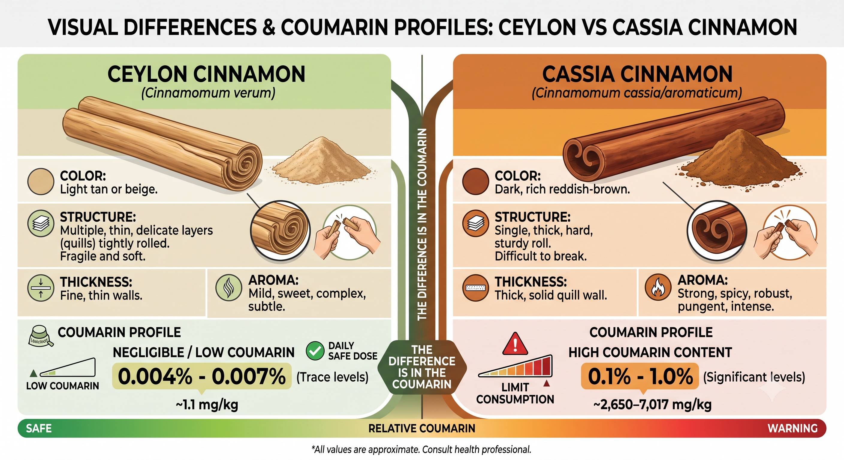
While cinnamon is highly regarded for improving insulin sensitivity and managing blood sugar, it can paradoxically trigger inflammation under specific conditions. The primary systemic risk stems from Cassia cinnamon, the common grocery store variety, which contains high concentrations of a compound called coumarin. When consumed regularly or in large therapeutic doses, coumarin can overwhelm the liver's metabolic pathways, leading to elevated liver enzymes and acute hepatic inflammation. Beyond liver toxicity, cinnamon can also incite localized inflammation; its volatile oil, cinnamaldehyde, can induce a Type IV delayed hypersensitivity reaction known as contact stomatitis - causing painful burning and swelling in the mouth - while concentrated doses can physically irritate the gastrointestinal lining. To safely harness the metabolic benefits of the spice without triggering these inflammatory responses, switching to Ceylon cinnamon ("true cinnamon") is the most effective strategy. Systemically, Ceylon contains only trace, negligible levels of coumarin (0.004% to 0.007%), making it safe for daily medicinal dosing, whereas Cassia contains significant levels (up to 1.0%) that require strict consumption limits to avoid toxicity. Visually and structurally, the two varieties are distinctly different: Ceylon features a light tan color with multiple thin, fragile, and tightly rolled layers, while Cassia is characterized by a dark reddish-brown hue and a single, thick, hard quill that is difficult to break.
(https://vitanetonline.com:443/forums/Index.cfm?CFApp=1&Message_ID=6645) Does Methylation Effect Active Vitamin D3 Levels in the body?
Date:
July 08, 2026 12:43 PM
Yes, methylation directly and significantly impacts how efficiently your body converts Vitamin D3 into its active, usable form. This relationship operates on two distinct levels: epigenetic DNA methylation (which acts as a volume knob for the conversion enzymes) and the cellular methylation cycle (which shares a profound reciprocal feedback loop with active Vitamin D). To see exactly where methylation interferes, it helps to look at the standard two-step activation pathway:
1. DNA Methylation (The Epigenetic "Dimmer Switch")DNA methylation is an epigenetic mechanism where methyl groups are attached to a gene's promoter region, typically silencing or "turning down" its expression.
2. The Systemic Methylation Cycle Feedback LoopWhile S-adenosylmethionine (SAMe) is not a direct chemical cofactor required for the hydroxylation reactions themselves (which rely on NADPH and the cytochrome P450 enzyme network), the systemic methylation cycle and Vitamin D share a massive reciprocal cross-talk mechanism.
The Vitamin D "Non-Responder": Is Your DNA Dimming Your Supplement’s Power? Yes, higher active Vitamin D3 levels consistently cause homocysteine levels to drop. Large-scale observational studies and gold-standard randomized controlled trials (RCTs) confirm a distinct, inverse relationship: as your Vitamin D status optimizes, circulating total homocysteine decreases. When active D3 binds to the Vitamin D Receptor (VDR), it triggers specific genetic and cellular actions that clear homocysteine through multiple pathways. The Biological MechanismsActive Vitamin D doesn’t just lower homocysteine through one pathway; it exerts a multi-pronged push to keep the toxic amino acid from accumulating.1. Upregulation of Methionine Synthase (MTR)While active D3 supports the alternative BHMT (betaine) pathway, recent molecular research highlights an even more direct impact on the primary, folate-dependent remethylation loop.
2. Safeguarding Enzyme Function via Oxidative Stress ReductionThe primary enzyme responsible for clearing homocysteine, Methionine Synthase, is incredibly sensitive to oxidative stress. Under high inflammation or oxidative conditions, the cobalt atom at the heart of its B12 cofactor becomes oxidized, completely disabling the enzyme and causing a major homocysteine backup.
3. Preserving Liver and Kidney FunctionThe alternative remethylation pathway, BHMT, operates almost exclusively in the liver and kidneys. If these organs experience structural or metabolic strain, their capacity to process homocysteine drops drastically. Active Vitamin D protects hepatic and renal tissue architecture, ensuring the local cellular machinery required for the betaine-homocysteine conversion remains functional.What the Clinical Trials ShowIn human trials, the drop isn't just theoretical - it's highly measurable.The Clinical Evidence: In double-blind, randomized, placebo-controlled trials, individuals with low baseline Vitamin D and elevated homocysteine were given high-dose Vitamin D3 (e.g., 50,000 IU weekly) over a two-month period. The treatment groups consistently showed statistically significant drops in total serum homocysteine, alongside reductions in body mass index (BMI) and systemic inflammatory markers, while the placebo groups saw no change. If someone is dealing with stubborn hyperhomocysteinemia (elevated homocysteine) and pushing hard on methylated B-vitamins (like methylfolate and TMG/betaine) without seeing their numbers budge, a hidden Vitamin D deficiency or a high-methylation genetic barrier at the CYP2R1 liver enzyme is very frequently the missing link. Summary: The relationship between Vitamin D3 activation and the body's biochemical pathways operates as a highly coordinated, reciprocal feedback loop. On an epigenetic level, DNA methylation acts like a cellular dimmer switch for the liver enzyme CYP2R1, which is responsible for converting raw D3 into its circulating form. When the promoter region of this gene is heavily methylated, enzyme production is suppressed, explaining why some individuals act as clinical "non-responders" who struggle to raise their blood levels even with high-dose supplementation. Once Vitamin D is successfully activated, however, it turns around and directly reinforces the systemic methylation cycle by binding to the Vitamin D Receptor (VDR) and upregulating the BHMT gene, which is vital for maintaining the body's universal methyl donor (SAMe) pool. Conversely, maintaining robust levels of active Vitamin D3 plays a direct, clinically proven role in forcing circulating homocysteine levels to drop. When active D3 binds to its receptor, it triggers a multi-pronged clearance strategy by activating the Nrf2 cellular defense pathway, which upregulates Methionine Synthase (MTR) - the primary enzyme that converts toxic homocysteine back into harmless methionine. Furthermore, by acting as a powerful systemic anti-inflammatory, active Vitamin D lowers the oxidative stress that would otherwise oxidize and disable the delicate B12 cofactors inside these clearance enzymes. This dual action of increasing enzyme production while shielding existing enzymes from environmental damage explains why clinical trials consistently show significant reductions in serum homocysteine when Vitamin D status is optimized.
(https://vitanetonline.com:443/forums/Index.cfm?CFApp=1&Message_ID=6644) Did you know that glutathione is not only great for liver health, but it also promotes beautiful, radiant skin?
Date:
December 07, 2023 12:12 PM
Did you know that glutathione is not only great for liver health, but it also promotes beautiful, radiant skin?Glutathione (GSH), often considered as an amino acid but actually a tripeptide, is an antioxidant primarily synthesized in the liver. Composed of cysteine, glutamic acid, and glycine, it plays a crucial role in the synthesis and repair of DNA and protein, as well as the synthesis of prostaglandins. With its involvement in various functions like amino acid transport, toxin and carcinogen metabolism, immune system function, prevention of oxidative cell damage, and activation of enzymes, it is undoubtedly the most important tripeptide in the body. While the benefits of supplementing with glutathione are numerous, two particularly compelling reasons are its positive impact on liver health and beautiful skin, which are the key focus of this article. However, before diving into the specifics of liver health and skin benefits, it's important to review the data on glutathione depletion and absorption. GSH depletion can occur due to various oxidative stressors such as radiation, v.infections, enviro toxins, household chemicals, heavy metals, surgery, inflammation, burns, septic shock, and dietary deficiencies of GSH precursors and enzyme cofactors. Additionally, research suggests that GSH levels tend to decline with age. The bioavailability of glutathione as a dietary supplement has encountered challenges in the past. Studies in the 1990s suggested that oral GSH might be inactivated by peptidases in the gut, as the levels of glutathione in the body did not seem to correlate with dietary intake, despite its presence in fruits, vegetables, and meats. Moreover, previous studies showed no significant increase in blood GSH levels when subjects were given high doses of 1,000-3,000 mg. As a result, alternative strategies like supplementation with NAC were used to boost GSH levels. In 2014, something interesting happened that changed the way we look at the bioavailability of GSH. A groundbreaking study published in the Journal of Agricultural and Food Chemistry shed new light on the old research. This study showed that GSH, when taken in its intact form as OPITAC, a yeast-derived glutathione by Kohjin/Mitsubishi, can actually be rapidly transported across intestinal epithelial cells. Once inside, it gets rapidly converted into oxidized glutathione (GSSG) and accumulates in red blood cells and the liver, with only a small presence in plasma. So, although the GSH was indeed absorbed, it didn't show up in blood plasma because it transformed into GSSG and stored in the red blood cells and the liver. The bottom line is, supplementing with GSH is an effective way to boost GSH levels in the body. This finding was further confirmed in another study that described how OPITAC, as a yeast-derived glutathione by Kohjin/Mitsubishi, is directly absorbed in its electrochemically reduced form in the intestine, then transported in the blood in bound forms, and eventually deposited into the liver in its reduced form. But here's where it gets even more significant. A six-month randomized, double-blinded, placebo-controlled trial involving 54 adults was conducted to investigate the effects of oral GSH supplementation (250 or 1,000 mg/day, as OPITAC glutathione, Kohjin/Mitsubishi) on GSH levels in various parts of the body, including blood, erythrocytes, plasma, lymphocytes, and exfoliated buccal mucosal cells. The results were astounding. After one, three, and six months, GSH levels in blood increased significantly compared to baseline in both dosage groups. At the six-month mark, GSH levels skyrocketed 30-35 percent in erythrocytes, plasma, and lymphocytes, and a mind-boggling 260 percent in buccal cells in the 1,000 mg group (P < 0.05). Even in the low-dose group, GSH levels in blood and erythrocytes increased by 17 and 29 percent, respectively (P < 0.05). This research clearly demonstrates that supplementation with GSH is not only effective for increasing GSH levels in the body but also for maintaining them. So, to sum it all up, the evidence speaks volumes - supplementing with GSH can have a profound impact on your body's GSH levels, and trust me, that's definitely a good thing! Liver HealthWhen it comes to our well-being, the liver is a true superhero. Let's dive into some fascinating details about this essential organ. Did you know that the liver is not only the largest reservoir of GSH (glutathione) but also a major site of GSH manufacture in the body? Pretty impressive, right? Special cells in the liver work tirelessly to synthesize GSH, which plays a crucial role in detoxification. Speaking of detoxification, the liver is a champion in this field. Its cells have sophisticated mechanisms to break down toxic substances, be it internal or external compounds. During the detoxification process, the liver attaches or conjugates the toxins to water-soluble substances. This attachment makes the toxic molecules more water-soluble, less harmful, and easier to eliminate via urine or bile. In fact, glutathione conjugation produces water-soluble mercaptates that are excreted via the kidneys, effectively detoxifying acetaminophen and nicotine. Isn't it amazing how this process helps our bodies get rid of harmful substances? But that's not all. Adequate levels of glutathione are crucial for the elimination of fat-soluble compounds, particularly heavy metals like mercury and lead. What's more, GSH serves as a cofactor for various peroxidase enzymes, aiding in the detoxification of peroxides generated from oxygen radical attacks on biological molecules. It also assists transhydrogenase enzymes in reducing oxidized centers on DNA, proteins, and other biomolecules. Talk about a multitasker! The practical significance of this liver superhero was demonstrated in a study involving workers exposed to lead. A group of five workers received GSH at 200 mg/day for 30 days, while five others served as the control group. The results were striking. The group receiving GSH showed a significant increase in ALA dehydratase activity (which is inhibited by lead) compared to the control group (p < 0.05). This indicates that GSH could be a valuable solution for treating patients with lead poisoning. So, let's take a moment to appreciate the remarkable liver and its incredible role in maintaining our health and well-being! Alcohol IntoxicationAlcohol consumption is widely recognized for its capability to induce hepatic steatosis, also known as fatty liver disease, and disrupt biomembranes due to hepatic lipid peroxidation. This can lead to various lifestyle-related diseases and even hepatic cirrhosis by diminishing hepatic physiological function. Nevertheless, animal studies have shown that hepatic damage caused by alcohol intoxication can be mitigated by glutathione (GSH), a powerful antioxidant found in cells. To further investigate the impact of GSH supplementation on the effects of alcohol intake, a human crossover comparative study was conducted. The study involved twenty healthy men and women who were grouped into three categories: placebo, 100 mg GSH (as OPITAC glutathione, Kohjin/Mitsubishi), and 30 mg curcumin. The study evaluated laboratory parameters, including breath alcohol concentration at different time intervals (20, 60, 120, and 180 minutes post-alcohol consumption) as measured by an alcohol checker. Additionally, subjective feelings were assessed through a questionnaire. During the study, all participants consumed whiskey in a quantity equal to their body weight multiplied by 1.25 mL, and were instructed to drink the entire sample within 10 minutes. The results revealed that the breath alcohol concentration in the group supplemented with GSH significantly decreased compared to the placebo and curcumin groups at 20 (p<0.01), 60 (p<0.01), 120 (p<0.05), and 180 (p<0.08) minutes post-consumption. Furthermore, the GSH group reported lower levels of "sleepiness," "headache," and "upset stomach" in the subjective feeling questionnaire. Importantly, the concentration of aspartate aminotransferase (AST), an indicator of alcohol-induced organ damage, was significantly lower in the GSH group after two months compared to the placebo group. The oral intake of GSH has demonstrated its effectiveness in reducing alcohol consumption-related stress and improving long-term hepatic function. These findings highlight the potential benefits of GSH supplementation in alleviating the detrimental effects of alcohol intoxication on the liver. Nonalcoholic fatty liver diseaseNonalcoholic fatty liver disease (NAFLD) is a condition characterized by the build-up of fat in the liver of individuals who consume little or no alcohol. Unfortunately, NAFLD is quite common, affecting nearly one-third of all American adults. Interestingly, it often presents without readily apparent signs or symptoms, sometimes resulting in complications, and can lead to liver inflammation and scarring as the fat accumulates. Additionally, NAFLD is typically associated with conditions such as insulin resistance, central obesity, reduced glucose tolerance, type-2 diabetes, and elevated triglyceride levels. Recognizing the substantial role glutathione (GSH) plays in phase 2 liver detoxification, a pilot trial was conducted to examine the therapeutic effects of GSH supplementation in patients with NAFLD. The trial included 29 individuals, and the patients were provided with daily oral supplementation of GSH at a dose of 300 mg (in the form of OPITAC glutathione, from Kohjin/Mitsubishi). The patients' clinical parameters were assessed before and after the GSH supplementation, and liver fat and fibrosis were quantified as well. The primary goal of the study was to determine any changes in alanine aminotransferase (ALT) levels. The results indicated a significant decrease in ALT levels following the GSH supplementation. Furthermore, triglycerides, non-esterified fatty acids, and ferritin levels also showed a reduction. This pilot study provides promising evidence for the potential therapeutic effects of oral glutathione administration, even at practical doses, in patients diagnosed with NAFLD. However, further investigation through large-scale clinical trials is necessary to validate its efficacy. In summary, NAFLD is a prevalent condition with potential serious consequences, but studies like the aforementioned pilot trial shed light on potential treatment options such as GSH supplementation. The findings demonstrate the need for continued research in order to provide more conclusive evidence and expand our understanding of NAFLD management. Beautiful SkinBy activating melanocytes in the skin, there is a notable increase in melanin formation, resulting in various blemishes such as freckles, pigmentation, and UV-induced skin spots, commonly known as age spots or liver spots. This is especially prominent after prolonged sun exposure and tanning. Age spots appear when melanin becomes concentrated or "clumped" in areas that have had years of frequent sun exposure. Luckily, there are materials like glutathione that can prevent or improve such pigmentation-related skin conditions. Another aspect to consider is skin pigmentation, wrinkles, and pores. In a study conducted with eight women in their 30s or early 40s, each supplemented with 100 mg/day of GSH (as OPITAC glutathione, Kohjin/Mitsubishi) for two months, their skin conditions were evaluated using the Robo Skin Analyzer. Several parameters were analyzed, including skin brightness, the amount and area of skin pigmentation, number of pores, and number of wrinkles under the eyes. It was observed that all subjects' skin brightness improved when measured on the second day of the study. Additionally, over the course of the two months, both the amount and area of skin pigmentation decreased, leading to an improvement in blemishes and pigmentation. Not only did glutathione exhibit a whitening effect, but it also reduced the number of wrinkles under the eyes and minimized pores. Furthermore, a randomized, double-blind, two-arm, placebo-controlled study was conducted with 60 otherwise healthy medical students. The purpose was to investigate whether supplementing with 500 mg of glutathione daily for four weeks would affect the skin melanin index compared to a placebo. Melanin indices were measured at six different sites on the body. The results demonstrated that melanin indices consistently decreased at all six sites in subjects who received glutathione after four weeks. The reductions were statistically significant compared to those who received the placebo at two sites: the right side of the face and the sun-exposed left forearm (p = 0.021 and 0.036, respectively). This improvement was likewise reflected in the reduction of UV spots. Importantly, both glutathione and placebo were well-tolerated. In conclusion, oral administration of glutathione leads to a lightening of skin color in the tested subjects. Skin LighteningSkin lightening is a process that is of interest to many individuals who seek to achieve a more even and radiant complexion. In recent studies, the use of a lozenge containing GSH 500 mg was explored as a means of skin lightening through an open-label, single-arm trial. The focus of this trial was to evaluate the buccal mucosa as a route for GSH administration and its potential in relation to skin lightening. It is worth noting that substances absorbed through the buccal route have the advantage of entering directly into the systemic circulation, effectively bypassing the gastrointestinal tract. The trial involved thirty Filipino females with Fitzpatrick skin types IV or V who received a daily glutathione-containing lozenge for eight weeks. The results from this trial demonstrated a significant decrease in melanin indices from baseline to endpoint. What is fascinating is that this visible change became evident in as little as two weeks. It is important to highlight that during this trial, there were no recorded serious adverse events, and the laboratory examination findings remained normal. Based on these findings, the researchers concluded that the lozenge containing glutathione was deemed safe and effective in lightening the skin of Filipino women. In addition to the aforementioned buccal route administration, another interesting approach that emerged from the studies is the topical application of GSH. A double-blind randomized clinical trial35 conducted in Yogyakarta, Indonesia, involved 74 healthy Indonesian women, with an average age of 33.3 ± 5.9 years, to explore the potential benefits of topical GSH. The trial subjects received supervised applications of facial wash twice a day, along with day cream containing sunscreen and night cream. The subjects were divided into three groups based on the active ingredients of the tested products, which included GSH (as OPITAC glutathione, Kohjin/Mitsubishi) at concentrations of 0.1 percent and 0.5 percent, and a control group without GSH. Throughout the trial, the effects of the tested products on skin color and pigmentation were measured using colorimetry with Chromameter Minolta for L. Compared to the baseline measurements, there were significant increases in lightness (L) detected as early as week 2 for the group using GSH at 0.1 percent concentration. Interestingly, this increase was significantly higher compared to the group using GSH at the higher concentration of 0.5 percent, as well as the group without GSH. It is important to note that hyperpigmented lesions also showed improvement, particularly in the group using GSH at 0.5 percent concentration, which displayed superiority compared to the other groups at week 8. In conclusion, the skin care products containing GSH at 0.1 percent and 0.5 percent concentrations were found to be effective in lightening facial skin. The findings from these studies shed light on the potential benefits of GSH in achieving skin lightening, either through buccal administration or topical application. It is worth noting that these studies focused on specific populations and more research is necessary to explore its effectiveness and safety across different skin types and ethnicities. In summary, the administration of
(https://vitanetonline.com:443/forums/Index.cfm?CFApp=1&Message_ID=6592) Can the Liver Be Repaired Once It Is Damaged?
Date:
April 29, 2022 11:33 AM
https://vitamins.vitanetonline.com/wp-content/uploads/liver-2934612_1920.png What is the liver and what does it do? The liver is a large, multi-functional organ that plays a vital role in the body. Its primary function is to filter the blood and remove toxins, but it also helps to regulate metabolism, store nutrients, and produce bile. The liver is divided into two main lobes, the right lobe and the left lobe. The right lobe is slightly larger than the left lobe and includes the quadrate lobe and caudate lobe. The left lobe contains the left lateral section and the medial section. The hepatic veins drain blood from the liver and pass it through the inferior vena cava to be returned to the heart. The hepatic arteries supply oxygenated blood to the liver tissue, while the portal vein carries nutrient-rich blood from the digestive organs. The bile ducts transport bile produced by the liver cells to the gallbladder, where it is stored until needed for digestion. The liver is a hardworking organ that performs many crucial functions in the body. By keeping it healthy, we can help to ensure that our entire body remains healthy and functioning properly. What can damage the liver and how does that damage occur? The liver is a vital organ that performs many important functions, such as filtering toxins from the blood and producing bile to help digest fats. However, the liver can be damaged by a number of different agents, including viruses, alcohol, and certain medications. Viruses such as hepatitis C and hepatitis B can cause inflammation of the liver, which can lead to scarring and eventually to liver failure. Alcohol abuse is also a major cause of liver damage, as it can lead to fatty deposits, inflammation, and scarring. In addition, certain drugs (such as acetaminophen) can cause toxic damage to the liver cells. If the liver is not functioning properly, it can result in a build-up of toxins in the blood, which can lead to serious health problems. Can the liver be repaired once it is damaged, and if so, how is that done? The liver is a amazing organ. It is responsible for so many important functions in the body, from filtering toxins to producing bile to breaking down fats. When it is damaged, it can often repair itself. This is because the liver has a high regenerative capacity. hepatocytes, which are the main type of liver cell, can divide and grow back quickly when necessary. In fact, as long as about 25% of the liver remains healthy, it can regenerate itself completely over the course of a few weeks. There are a number of ways to damage the liver, such as through alcohol abuse or viral infections, but fortunately there are also ways to help it repair itself. Eating a healthy diet, getting enough exercise, and avoiding toxins, take liver supporting supplements like milk thistle and NAC can all support liver health and regeneration. How can people prevent their livers from being damaged in the first place? The liver is a vital organ that plays an important role in metabolism. However, the liver can also be easily damaged by alcohol, drugs, prescriptions, and environmental toxins, which are in the foods we eat. To help prevent liver damage, it is important to avoid excess alcohol consumption or avoid it all together, take medication as directed and if you are on medications that cause liver damage to discuss alternatives that do not harm the liver, and eat a healthy diet along with taking liver supporting supplements. Additionally, it is important to get regular exercise, as this helps to keep the liver healthy and functioning properly. By taking these steps, people can help to prevent liver damage and keep their organs healthy. Are there any lifestyle changes or supplements that can help improve liver function even if it has been damaged already? Liver damage is a serious concern for many people, as it can lead to a number of health problems. While there is no guaranteed way to reverse liver damage, there are some lifestyle changes and supplements that may help improve liver function. One potential change is to limit alcohol consumption, as alcohol is known to be damaging to the liver, mixing alchol and drugs can enhance the progress of liver damage. Additionally, eating a healthy diet and exercising regularly can help improve overall liver function. There are also a number of herbal supplements that are thought to be beneficial for the liver, such as milk thistle and dandelion root. However, it is important to speak with a healthcare provider before starting any new supplement, as some may interact with other medications. What are the possible consequences of not repairing a damaged liver? Liver damage can have serious consequences if left untreated. Depending on the cause, there may be a variety of negative effects that result from an injured liver. For example, if the damage is due to excessive alcohol consumption, then scarring and inflammation of the liver tissue can interfere with its ability to filter toxins out of the blood. This can lead to a host of potentially dangerous health problems, including nausea, chronic fatigue, jaundice, and cirrhosis. You must address the cause before change for the good can happen. In addition, unresolved inflammation can increase one's risk of developing more serious conditions such as cancer or heart disease. To prevent these possible consequences, it is important to seek medical attention as soon as possible after suffering from liver damage. This will allow doctors to properly diagnose the issue and develop an appropriate treatment plan. Addressing the cause is always preferable over simply treating symptoms alone. Ultimately, taking action now can help you protect your health in the future. How can people find out if their livers need repair and where they should go for help if that is the case? When it comes to liver health, there are a number of ways to find out if your liver needs some repairs. One method is to get tested for elevated liver enzymes, which are indicative of problems with the organ. You can also watch out for certain symptoms, such as fatigue, bloating or skin discoloration. If you notice any of these markers, you may want to consult with your doctor or a specialist in liver disease, who can help determine the best course of action. Depending on the underlying cause of your liver problem, treatment options may include changes to your diet and exercise habits, medications or even surgery in extreme cases. Whatever route you choose, it is essential that you address any issues with your liver as soon as possible in order to protect your overall wellbeing. Generally, symptoms only show when liver function has dropped significantly. Liver damage can often go undetected until it has reached a advanced stage. This is because the liver has an incredible capacity for regeneration and can continue to function even when up to 75% of it has been damaged. As a result, symptoms of liver disease often only become apparent when the organ has been severely compromised. By that point, the damage may be irreversible and serious complications may have developed. Therefore, it is important to be aware of the early signs of liver damage so that treatment can be started as soon as possible. These include fatigue, weight loss, abdominal pain, and yellowing of the skin and eyes. Fortunately, Supplements like Milk thistle and NAC may help the body assist the liver in its repair. The liver is a hardworking organ that is constantly exposed to toxins. As a result, it is susceptible to damage. Fortunately, supplements like milk thistle and NAC may help the body assist the liver in its repair. Milk thistle contains silymarin, a compound that helps to protect the liver from damage. NAC is an amino acid that helps to remove toxins from the liver. NAC also helps to replenish glutathione, a substance that plays a key role in liver detoxification. Together, these supplements can help to keep the liver healthy and functioning properly. Whether you are looking for preventative measures to protect the liver or have an existing issue, it can never hurt to take either milk thistle, NAC, or both to support healthy liver function.
(https://vitanetonline.com:443/forums/Index.cfm?CFApp=1&Message_ID=6396) Pu-erh tea has hepatoprotective and antioxidant properties
Date:
May 14, 2019 04:25 PM
Yunnan Tasly Deepure Biological Tea Group Company recently collaborated with scientists from Tianjin University of Commerce on research into the health benefits of pu-erh tea. The research involved rats on a high fat diet, and discovered that pu-erh tea can help defend your liver against oxidative stress and high glucose levels. Pu-erh tea also appears to be able to protect rats from weight and body fat increases and prevent lipid peroxidation. These results suggest pu-era tea has great potential for preventing fatty liver disease. Key Takeaways:
"The results showed that the treatment with Pu-erh tea extract prevented the increases of body weight and fat index, reduced oxidative stress, and inhibited lipid peroxidation — all of which contributed to the protection of the liver in rats." Read more: https://www.naturalnews.com/2019-04-16-pu-erh-tea-has-hepatoprotective-and-antioxidant-properties.html
(https://vitanetonline.com:443/forums/Index.cfm?CFApp=1&Message_ID=6317) Here's how the body prevents helpful bacteria from causing disease
Date:
February 11, 2018 03:59 PM
The body is pretty amazing in how it can detect foes. The system is literally called "friend/foe" and it is the immune system's response to all bacteria. Researchers examined mice for a particular strand of bacteria known as Helicobacter hepaticus. This invader caused a response from T cells and immediately was acted upon as "foe." Specifically Th17 were the T cells that were able to kill the bacteria, but they have another which shuts this cell down when finding friendly ones, not causing the inflammation chain that it normally does. Key Takeaways:
"Certain bacteria, such as those from the Helicobacter family, are generally considered helpful but can cause disease when genetic or environmental factors alter the normal balance." Read more: https://www.breakingnews.ie/world/heres-how-the-body-prevents-helpful-bacteria-from-causing-disease-827150.html
(https://vitanetonline.com:443/forums/Index.cfm?CFApp=1&Message_ID=5526) What is Bioperine and How Does It Help with Absorption of Vitamins
Date:
April 21, 2011 03:14 PM
Get more from your food with Bioperine.Bioperine is a patented form of an alkaloid found in black pepper. It is derived entirely from piperine, an organic compound responsible for the spicy taste of black pepper and long pepper. Piperine has been noted for its thermogenic properties believed to speed up the absorption metabolism of digested foods. Recent studies have discovered that it also interferes with the release of enzymes that govern the bioavailability of drugs and supplements. By so doing, it enhances the rate of absorption of vitamins. The human body has a complex mechanism of controlling the substances that get in and out of systemic circulation. The first pass occurs in the alimentary canal, where gastrointestinal enzymes break down substances into smaller compounds. It is believed that a very small percentage manages to undergo intestinal absorption after digestion. In general, this is the part where constituents of supplements remain undigested and instead enter the colon together with waste materials. The liver plays a central role to the metabolism of drugs and most bioactive compounds. The compounds that pass the intestinal walls and enter the hepatic portal system, a group of veins that direct blood and other compounds from the gastrointestinal tract to the liver, is further metabolized inside the liver. Anything that the body considers foreign is sent to the kidneys and easily excreted through the urine. This is the reason why bioavailability is significantly reduced after ingestion. Counteracts Effects of Enzymes Bioperine is the only compound known to interact with enzymes that controls the metabolism of foreign materials within the intestinal epithelium. P-glycoprotein is released in the digestive tract to deal with drugs and xenobiotics found in our diet. Vitamins are no exception to the tightly regulated process of absorption in the intestines. These enzymes transport digested compounds to the liver. It has been observed that piperine appears to reduce the expression of p-glycoprotein in the alimentary canal and other parts of the body. Proponents believe that bioperine, a purer form of piperine, is capable of counteracting the effects of the enzyme within the intestinal epithelium, making it easier for vitamins and supplements to enter the hepatic portal system. Increases the Rate of Metabolism Not all compounds that undergo the first-pass effect interact with the cells and tissues they are supposed to act on. If they should have an effect on any cell, they are still subjected to the actions of enzymes specialized for the expulsion of xenobiotics found in the systemic circulation and the rest of the body. For example, CYP3A4 removes foreign materials from the cells and facilitates their excretion. Bioperine is touted to induce thermogenesis and stimulate cellular activities. By so doing, the effects of bioactive compounds are achieved while they remain inside the cells and tissues. The thermogenic properties of bioperine influence the rate of metabolism of digested compounds, including vitamins, minerals, and components of herbal preparations. Note that it can increase the update or prescription medications as well, so caution should be observed when consuming bioperine with medications. If you want to give your body a nutrient boost, add bioperine to your supplement regiment to boost absorption.
(https://vitanetonline.com:443/forums/Index.cfm?CFApp=1&Message_ID=2280) Cascara sagrada can help with constipation
Date:
August 25, 2010 02:18 PM
Cascara sagrada and ConstipationCascara Sagrada was introduced to the Spanish explorers by Native Americans when they complained of problems with constipation. The Spanish then gave this herb its name, cascara sagrada, which means sacred bark and used it as a natural laxative. The Spanish explorers brought the herb back with them to Spain when they returned. In 1877, this herb was admitted to the U.S. Pharmacopoeia and is still included as an official medicine to this day. Cascara sagrada is believed to be one of the best herbs for treatment of chronic constipation. This is because it enhances the peristaltic action that takes place in the intestines and increases the secretions of the stomach, liver, and pancreas. This herb is also helpful in relieving hemorrhoids because of its nonirritating nature and softening action on the stool. Cascara sagrada is found in many over-the-counter preparations that are used for the intention of relieving constipation. This is because it acts on the large intestine to increase the muscular activity of the colon. The anthraquinones that are found in cascara sagrada are believed to encourage intestinal contraction. This herb is used to restore natural bowel movement without griping and to restore tone to the bowel.
Additionally, an element in cascara sagrada is known as quinine emodin. This element is being studied for its usefulness in treating lymphocytic leukemia and Walker carcinosarcoma tumor Cascara sagrada can be used often, as it is not considered to be addictive. Preparations should be made from bark that has been aged for a least a year before use. This must be done because fresh bark is poisonous and can cause nausea and extreme griping on the intestinal system. Cascara sagrada can be found in various forms including capsules, liquid extracts, and dried bark. Although it tastes bitter, the dried bark can be made into a tea. A typical dosage of cascara is a 300 mg capsule which is taken in the early evening to stimulate a bowel movement in the morning. The laxative effect usually occurs 6 to 12 hours after cascara is taken. Products containing cascara sagrada should not be used for more than eight or ten days. Those women who are pregnant and nursing should avoid this herb, as well as people with intestinal blockage, undiagnosed stomach pain, or symptoms that may indicate appendicitis. People with diarrhea, inflammatory bowel disease, or intestinal ulcers should not take this herb. Additionally, children younger than twelve with constipation should not be treated with cascara sagrada. The bark of cascara sagrada should be used to provide alterative, antineoplastic, antispasmodic, hepatic, lithotriptic, and purgative properties. The primary nutrients found in this herb are calcium, chlorine, iron, magnesium, manganese, niacin, phosphorus, potassium, selenium, silicon, sodium, and vitamins A, B-complex, and C. Primarily, cascara sagrada is beneficial in dealing with colon problems, constipation, gallbladder problems, gallstones, gas, gastric disorders, hemorrhoids, intestinal problems, liver disorders, and worms. Additionally, this herb is extremely helpful in treating colitis, coughs, croup, dyspepsia, gout, indigestion, insomnia, jaundice, excessive mucus, pituitary problems, and spleen ailments. For more information on the many beneficial effects provided by cascara sagrada, please contact a representative from your local health food store. Always choose name brands of this wonderful herbal laxative to ensure quality and purity and gentleness of its properties on the intestinal tract.
(https://vitanetonline.com:443/forums/Index.cfm?CFApp=1&Message_ID=2180) Royal Jelly As An Anti-Aging Supplement
Date:
February 27, 2010 11:45 AM
Gelatin is another component of royal jelly and one of the primary precursors of collagen. It is a potent anti-aging compound that helps to keep the youthful, while supporting the organs, glands, and muscular systems. It contains vital fatty acids, sugars, sterols, phosphorus compounds, and acetylcholine. Acetylcholine is essential in order to properly transmit nerve impulses. It is also important for the proper functioning of the endocrine system. An insufficient amount of acetylcholine often leads to a susceptibility to a variety of nerve disorders such as Alzheimer’s disease, Parkinson’s disease, and multiple sclerosis. Royal jelly has many properties. Among which include: antibacterial, antiviral, antibiotic, tonic, nutritive, and anti-aging. It is very beneficial to the immune, cardiovascular, endocrine, integumentary, nervous, reproductive, cellular, skeletal, hepatic, and respiratory systems. Although aging is inevitable, the process can be slowed significantly with supplements. Researchers in Argentina have been working to document the ability of royal jelly to both slow down tissue deterioration and even reverse it. For example, the story of Noel Johnson should be noted, as he experienced of rebirth of health at age eighty. In 1964, at age sixty-five he was refused life insurance because of a weak and damaged heart. At this point he was also cautioned to restrict his physical activity in order to prevent death. In 1989, at age ninety, Johnson discussed the book he wrote, The Living Proof… I Have Found the Fountain of Youth, which claims that his use of bee products, along with a solid nutritional program, is responsible for changing his life. Despite the additional exercise and diet changes, Johnson attributes most of his vitality to the bee products he used. He claims that he discovered bee’s gift at age seventy, as the foods restored his manhood, brought him to full vigor and sexual potency, and continue to nourish every cell in his body. Johnson claims to be improving in every way without spending any money on medicine. Royal jelly has been found to help with many conditions. Some of these include menopause, impotence, infertility, chronic fatigue, skin blemishes, wrinkles, immune system problems, viral and bacterial infections, endocrine disorders, hormonal balance, cardiovascular disease, weight control, inflammation, liver ailments, cancer, arthritis, memory, depression, Parkinson’s disease, diabetes, asthma, and mental exhaustion. In order to obtain the best results when using this, or any bee product, it is important to consult your health care provider before beginning any regimen. For more information on the many beneficial effects provided by royal jelly, please feel free to consult a representative from your local health food store with questions. Always buy Name brands like Premier One Royal Jelly to ensure quality and purity of the product you purchase.
~video~
(https://vitanetonline.com:443/forums/Index.cfm?CFApp=1&Message_ID=2128) Gentain Herb Can Sooth The Digestive Tract
Date:
December 17, 2009 04:23 PM
Gentian was used as a stomach tonic and aid in digestion in Ancient Rome. This herb is native to Europe and western Asia. Generally, gentian was consumed as a tea or alcoholic beverage. Gentian was an official drug in the Untied States Pharmacopoeia from 1820 to 1955 and was used as a gastric stimulant. At one point in time, this herb was used and acclaimed by medical science as being very beneficial for mankind. Gentian was used to reduce fevers by cooling the system. It contains a bitter principle, which is known as amarogentin, which stimulates the glands, and includes the adrenals and thyroid. This herb helps in the production of bile, which can have a positive effect on the liver and gallbladder. Gentian is also used to clean the bowels, stimulate the pancreas, stimulate circulation, aid in the digestive process, and help with female problems. This herb has historically been used to treat wounds and been taken internally for inflammation from arthritis, jaundice, and a sore throat. Gentian is considered to be a great herb for strengthening the entire body and for use as tonic when it is combined with other herbs. German scientists have performed studies which confirm that this herb is useful as a digestive aid. Herbal bitters, including gentian, are recommended for the treatment of indigestion. The bitter taste receptors located in the tongue are known for their ability to stimulate the digestive processes by increasing the flow of gastric juices and bile. The alkaloid found in gentian, gentianine, has been shown to contain anti-inflammatory activity in animal studies.
Internally, gentian is used as a liver tonic, to treat loss of appetite, digestive problems, flatulence, and insufficient production of gastric juices and saliva. This herb is responsible for stimulating the taste buds and promoting the flow of saliva, gastric juices, and bile. Because of this, gentian can be used in cases of anorexia, and in homeopathic medicine as well. There are no external uses or aromatherapy and essential oil uses for gentian. It should be noted that gentian should not be used by those with gastric or duodenal ulcers. The root of the gentian plant is used to provide alterative, anthelmintic, anti-inflammatory, antiseptic, antispasmodic, blood purifier, emmenagogue, febrifuge, hepatic, sialagogue, stimulant, stomachic, and vulnerary properties. The primary nutrients provided by gentian include inositol, iron, manganese, niacin, silicon, sulfur, vitamins F and B-complex, and zinc. Primarily, gentian is extremely beneficial in treating loss of appetite, poor circulation, gastric disorders, indigestion, jaundice, and liver disorders. Additionally, this herb is very helpful in dealing with anemia, blood impurities, colds, constipation, stomach cramps, diarrhea, dysentery, fevers, gas, gout, heart burn, absence of menstruation, nausea, spleen ailments, urinary problems, worms, wounds, and yeast infections. For more information on the many beneficial effects provided by gentian, please contact a representative from your local health food store with questions.
(https://vitanetonline.com:443/forums/Index.cfm?CFApp=1&Message_ID=2117) Horse Radish
Date:
October 28, 2009 11:39 AM
Horseradish has been cultivated since ancient times. The Delphic Oracle in Greek mythology told Apollo that horse radish was worth its weight in gold. Horseradish was known in Egypt by 1500 BC and has been used by Jews from Eastern Europe traditionally in Passover. The plant is discussed by Cato in his treatises on agriculture. It is thought that horseradish is the plant known as Wild Radish by the Greeks. Both the root and leaves of the horseradish plant were used as a medicine during the Middle Ages, with the root used as a condiment on meats in Germany, Scandinavia, and Britain. This herb was taken to North American during Colonial times. It is not certain as to where the name horseradish come from. Some believe that it derives by misinterpretation of the German Merettich as mare radish. Others think the name comes from the coarseness of the root. The common thought in Europe is that it refers to the old method of processing the root called hoofing, in which horses were used to stamp the root tender before grating it.
For at least two thousand years, horseradish has been cultivated. It was brought to America by early settlers and used to treat conditions such as pain from sciatic, colic, and intestinal worms. Horseradish provides antibiotic action that is recommended for respiratory and urinary infections. The volatile oil in horseradish has the ability to work as a nasal and bronchial dilator. Internally, it has been used to clear nasal passages, alleviate sinus problems, help with digestion, work as a diuretic, aid with edema and rheumatism, and cleanse various body systems. Also, horseradish has been used to stimulate digestion, metabolism, and kidney function. The root of the horseradish plant can be used to provide antibiotic, antineoplastic, antiseptic, bitter, caminative, diaphoretic, digestive, diuretic, expectorant, hepatic, parasiticide, mild purgative, rubefacient, sialagogue, stimulant, and stomachic properties. The primary nutrients found in this herb are calcium, iron, phosphorus, sodium, and vitamins A, B-complex, and P. Primarily, horseradish is extremely beneficial in dealing with loss of appetite, circulation, coughs, edema, excessive mucus, sinus problems, internal and skin tumors, and worms. Additionally, this herb is very helpful in treating arthritis, asthma, bronchitis, congestion, gout, jaundice, kidney problems, irritated membranes, neuralgia, palsy, rheumatism, skin conditions, water retention, and wounds. For more information on the many beneficial effects provided by horseradish, please feel free to contact a representative from your local health food store with questions.
(https://vitanetonline.com:443/forums/Index.cfm?CFApp=1&Message_ID=2107) Goldenseal Root
Date:
October 06, 2009 01:22 PM
Traditionally, goldenseal has been used for many different conditions. Among these are boosting the glandular system, hormone imbalance, congestion, inflammation, female problems, infection, bronchitis, menstrual problems, catarrh of the bladder, gastritis, ulcers, bowel stimulation, antiseptic, and as an immune system builder. Those with low blood sugar or pregnant women should not use this herb.
Recent studies have determined that goldenseal is beneficial in fighting viruses and infections. This herb contains the alkaloids hyrastine and hyrastinine, which possess strong astringent and antiseptic benefits on the mucous membranes. The berberine that is found in goldenseal, and can also be found in barberry, Oregon grape, and goldthread, is effective in fighting infections of the mucous membranes, which includes the mouth, throat, and sinuses. It has also been found to kill toxic bacteria in the intestinal tract like giardiasis, which is found in streams of North America. Goldenseal can help to relieve diarrhea in cases of giardiasis, amoebiasis, or other gastrointestinal infections. The alkaloid content of goldenseal gives it its antibiotic properties. Goldenseal has a long history of use for fighting both colds and flu viruses. The berbine content is effective as a natural antibiotic and immune stimulant. The herb may also help to prevent candida infection which is the result of antibiotic use. Goldenseal is thought to help strengthen the immune system and may work by increasing the blood supply to the spleen. This enables the spleen to function and release compounds which are known to improve immune function. Some herbalists in England consider goldenseal to be the wonder remedy for digestive problems. This herb is recommended for use after the onset of a cold instead of as preventative action. For this reason, it is often found in cold remedy combinations.
The rhizome and root of the goldenseal plant are used to provide adaptogen, alterative, anthelmintic, antibiotic, antiperiodic, antiseptic, cholagogue, emmenagoggue, hepatic, nephritic, stomachic,, and mild purgative properties. The primary nutrients found in this herb are calcium, copper, iron, manganese, phosphorus, potassium sodium, vitamins A, B-complex, C, E, and F, and zinc. Primarily, this herb is extremely helpful in treating bronchitis, poor circulation, colds, colitis, colon problems, coughs, diarrhea, eye infections, gonorrhea, gum disease, hemorrhages, hemorrhoids, infection, inflammation, intestinal problems, kidney problems, liver disorders, excessive menstruation, membrane infections, mouth sores, nosebleeds, and sore throat. Additionally, this herb is very helpful in dealing with allergies, hay fever, asthma, Bright’s disease, burns, chicken pox, constipation, earaches, eczema, fever, flu, gallbladder problems, gastric disorders, gastritis, glandular problems, heart conditions, herpes, membrane irritation, nausea, nervous disorders, ringworm, skin disorders, spleen ailments, tonsitilits, and urinary problems. For more information on the many beneficial effects provided by goldenseal, please feel free to contact a representative from your local health food store.
(https://vitanetonline.com:443/forums/Index.cfm?CFApp=1&Message_ID=2098) Milk Thistle And Silymarin
Date:
August 12, 2009 12:44 PM
In Europe, milk thistle was used as a well-known remedy for liver problems and also as a digestive aid. Pliny the Elder, the early Roman writer, explained how the juice of milk thistle mixed with honey was used for carrying off bile. Gerard, an herbalist, said in 1597 that milk thistle was one of the best remedies for liver-related diseases. Milk thistle was also given to nursing mothers to improve milk production. The liver is an extremely important organ that is found in the body. It is responsible for filtering toxic material from the body, which prevents accumulation that can lead to disease and death. Often, the vital functions of the liver are overlooked. Nevertheless, they are extremely important. It is essential to keep the liver working properly. Milk thistle has been proven to be very beneficial for liver function. Milk thistle extract has been shown in observations to help reverse both acute and chronic liver problems, including cirrhosis and viral hepatitis. The antioxidant properties may be due to the bioflavonoid content. Milk thistle has also been found to help heal the liver from damage that occurs from alcohol toxicity. It has been used to treat many different liver ailments. Among these are fatty liver disorders, chronic hepatitis, inflammation of the bile duct, hardening of the liver, and cirrhosis. Milk thistle is also thought to actually help liver regeneration when part of the liver is removed. Milk thistle possesses a variety of compounds which are known as Silymarin. This includes silybin, silydianin, and silychristin. All of these substances are known to protect the liver against some toxins, which helps to increase the function of this important organ. Silybin is used as an antidote to the deathcape mushroom, which is known to destroy liver cells. The deathcap species is one of the most toxic of liver poisons and has a death rate of up to fifty percent. A study with sixty patients suffering from deathcap poisoning treated with silybin produced incredible results. None of the participants died. Silymarin is an effective remedy if it is administered within forty-eight hours of ingestion. This compound seems to occupy the receptor sites to protect the cell membranes. Not only does it work to treat serious liver conditions, but it also prevents damage from occurring. Silymarin contains amines which are known to help stimulate the production and flow of bile. This compound has been found in studies to work exclusively on the kidneys and liver. Milk thistle has also been shown to stimulate the synthesis of protein. The seeds of the milk thistle plant are used to provide alterative, antioxidant, galactagogue, hepatic, and stimulant properties. The primary nutrients found in this herb are bioflavonoids. Primarily, milk thistle is extremely beneficial in treating cirrhosis, hepatitis, jaundice, kidney problems, and liver disorders. Additionally, this herb is very helpful in dealing with alcoholism, appetite loss, high blood pressure, boils, chemotherapy, depression, epilepsy, fatty deposits, gas, heartburn, heart problems, hemorrhages, hypoglycemia, indigestion, absent lactation, menstrual symptoms, effects of radiation, skin diseases, varicose veins, and the effects of toxins.
(https://vitanetonline.com:443/forums/Index.cfm?CFApp=1&Message_ID=2052) Oregon Grape Extract
Date:
August 11, 2009 01:21 PM
Oregon grape is an evergreen shrub that is related to the barberry plant. The Oregon grape is not closely related to grapes, but it gets its name from the purple clusters of berries. The color and slightly duster appearance is similar to that of grapes. Often, it is referred to as the “tall Oregon grape” in order to distinguish it from the “creeping Oregon grape” and “dwarf Oregon grape.” The Oregon grape grows approximately one to five meters tall. It has leathery leaves that resemble holly and stems and twigs that have a thick, corky appearance. The flowers, which grow in late spring, are a bright yellow color. This plant is often used in landscaping similarly to barberry. The plant is suited for low-maintenance plantings and loose hedges. This plant is resistant to summer drought, tolerates poor soils, and does not create excessive leaf litter. The berries of the Oregon grape attract birds. The purplish-black fruits found on the Oregon grape plant are quite tart and contain large seeds. Sometimes, they are used locally and mixed with Salal to make jelly. The fruit is bitter and generally not eaten unless it is sweetened first. The leaves of the Oregon grape are holly-like and resist wilting. For this reason, the foliage is often used by florists for greenery. Additionally, the inner bark of the larger stems and roots yield a yellow dye. The Oregon grape plant grows natively on the North American west coast from British Columbia to northern California. It is also the state flower of Oregon. In some areas outside of its native range, this plant has been classified as an invasive exotic species that may displace native vegetation. Oregon grape tonics were first introduced as a medicinal remedy in the late nineteenth century. The herb was marketed as a blood purifier. Oregon grape is well known for the treatment of skin diseases that are caused by toxins in the blood. This is because it stimulates the action of the liver. It is also one of the best blood cleansers. This herb is also mildly stimulates thyroid function. This herb aids in the assimilation of nutrients, promotes digestions, and is a tonic for all glands. The rhizome and root of the Oregon grape plant are used to provide alterative, antiseptic, blood purifier, cholagogue, hepatic, nephritis, nutritive, and mild purgative properties. The primary nutrients found in this herb are copper, manganese, silicon, sodium, vitamin C, and zinc. Primarily, Oregon grape is extremely beneficial in treating acne, blood conditions, blood impurities, eczema, jaundice, liver disorders, psoriasis, and staph infections. Additionally, this herb is very helpful in dealing with rheumatoid arthritis, bronchitis, chronic constipation, hepatitis, herpes, intestinal problems, kidney problems, leucorrhea, lymphatic problems, rheumatism, lack of strength, syphilis, uterine problems, and vaginitis. In order to obtain the best results when supplementing with this, or any herb, it is important to consult your health care provider before beginning any regimen. For more information on the many beneficial effects provided by Oregon grape, please feel free to consult a representative from your local health food store with questions.
(https://vitanetonline.com:443/forums/Index.cfm?CFApp=1&Message_ID=2050) Barberry
Date:
May 13, 2009 12:39 PM
The barberry plant is a shrub that has gray, thorny branches. This shrub can grow up to nine feet tall. The flower of the barberry plant are bright yellow and bloom between the months of April and June. These flowers then become dark, drooping bunches of red berries in the fall. The use of barberry dates back approximately three thousand years, originating in China in India where it was used for the treatment of diarrhea and intestinal infections. The barberry plant was used by Native Americans for treating liver conditions like jaundice. Additionally, Egyptians mixed the berries of the plant with fennel seed to protect themselves from the plague. Barberry is made up of an alkaloid known as berberine, which can also be found in other medicinal herbs such as goldenseal and Orgeon grape. The therapeutic effects of barberry can be attributed to its berberine content. Studies have concluded that berberine contains properties that are effective against a wide variety of bacteria, viruses, and fungi. These studies also found that berberine was much more effective in treating some bacteria than even a strong antibiotic. Other studies have found that barberry has the potential to kill microorganisms including staphylococci, streptococci, salmonella, Giardia lamblia, Escherichia coli, shigella, and Candida albicans. The berberine in barberry has been noted to contain antidarrheal properties. This alkaloid is also recommended for stimulating the immune system. The effects of barberry include helping against cancer, liver problems, kidney problems, coughs, cholera, diarrhea, fever, inflammation, hypertension, and tumors. Barberry has also been recommended to increase bile secretions and stimulate the appetite. This herb may also help in cases of anemia and malnutrition. Barberry stimulates bile production for liver problems and also dilates blood vessels to lower blood pressure. Barberry is used in easing inflammation and infection of the urinary, gastrointestinal, and respiratory tracts, as well as candida infections of the both the skin and vagina. Barberry extract has also been shown to improve symptoms that are associated with certain skin conditions, such as psoriasis. However, more research is still needed on determining the reliability of these findings. Barberry is shown to be an extremely effective treatment for diarrhea. A few studies have found that barberry is able to improve symptoms faster than antibiotics. This is possibly because of its astringent properties. However antibiotics are still thought to be more effective at killing bacteria in the intestines. For this reason, it is best to use barberry to ease symptoms, along with a standard antibiotic, as bacterial diarrhea can have extremely serious consequences. The bark, root, and berries of the barberry plant are used to provide alterative, antibacterial, antineoplastic, antiseptic, aromatic, astringent, blood purifier, cholagogue, diuretic, hepatic, hypotensive, purgative, and stomachic properties. The primary nutrients provided by this herb include iron, magnesium, phosphorus, and vitamin C. Primarily, barberry can be beneficial in dealing with loss of appetite, high blood pressure, impurities in the blood, candidiasis, constipation, diarrhea, dysentery, fevers, indigestion, infections, jaundice, liver disorders, pyorrhea, and sore throat. However, this herb is also extremely helpful in dealing with anemia, arthritis, boils, breath odor, cholera, gallstones, heart problems, heartburn, hemorrhages, itching, kidney problems, migraines, rheumatisms, ringworm, and skin conditions. For more information on barberry or to make a purchase, along with its many beneficial effects, feel free to contact a representative at your local health food store.
(https://vitanetonline.com:443/forums/Index.cfm?CFApp=1&Message_ID=2006) Milk Thistle May Help With Cirrhosis, Gallstones, and Hepatitis Liver Problems
Date:
November 14, 2007 12:34 PM
Milk thistle, botanically Silybum Adans, is a member of the daisy family that is native to the Mediterranean, the Middle East and North Africa but has also been introduced to California and parts of the eastern side of the USA, and can help with problems such as cirrhosis, gallstones and hepatitis. Although it has been recorded as being used in medieval times as a tonic for the liver, it is only relatively recently that its chemical components have been investigated. Analysis indicated the presence of hitherto unknown flavonoids which were given the name of silymarin. In general, flavonoids are strong antioxidants found in many fruits and vegetables that eat up the free radicals that cause so much damage to our bodies. Free radicals are very unstable molecules that are generated through pollution, such as smoking, car fumes, perticides etc, and that destroy body cells accelerating aging. They also oxidise the low density lipids (LDL) that carry water-insoluble cholesterol through the blood to the arteries where it is needed to repair damage, resulting in excess deposition and the atherosclerosis that can cause strokes and heart disease. Antioxidants mop these up like a sponge, and are some of the healthiest types of molecules that we can consume. The silymarin group of flavonoids are particularly attracted to the liver where they act as antihepatoxic agents that prevent the liver from becoming poisoned. But why should the liver be poisoned I can hear you ask, and what are the poisons involved? Can we avoid them? It is a very relevant question, and one that will make you wonder, every time you leave your home, especially if you live in a big city or an industrialized area. Smoking cigarettes, working with volatile organic compounds that you can breathe in, such as paint and printing ink solvents, the exhaust fumes of cars and diesel engines, factory chimneys belching out tons of smoke, analgesics such as paracetamol, pesticides on your fruit and vegetables that you have failed to wash off, alcohol, etc, etc, etc… All of these have to be removed from your body or you will die, awash with all these poisons that you have ingested, some deliberately and some not. The organ that does this is your liver: the powerhouse chemical plant of your body that carries out millions of biochemical reactions every day. Your liver converts all of these poisons into molecules that can be flushed away through your body naturally. However it places great stresses upon it, and even your liver needs a rest sometimes, or even just a little rejuvenating tonic would keep it happy. Milk thistle has been used for just that purpose, especially when the liver has been toxified with excess alcohol, pesticide poisoning or even hepatitis. The silymarin initially coats the cells of the liver by binding to the cell membranes of the cell walls, so that the toxins are hindered from entering the cells. Its antioxidant properties then neutralise any free radicals present that are causing the damage to the liver cells. It also helps to stimulate the production of proteins to help the healing process, and reduces the fibrosis that is the development of fibrous masses outside the liver cells caused by damaged cells excreting materials such as collagens outside the cells into the general body of the liver. Finally, milk thistle helps to prevent the activity of the immune system in causing inflammation of the damaged cells. Silymarin acts very specifically on the liver, and is often used by physicians in the treatment of such liver conditions as jaundice, hepatitis, liver cancer and cirrhosis. In addition to its own effects it appears that it stimulates the production of glutathione that is also a very powerful natural tripeptide antioxidant produced by the body when it is under oxidative stress. Its effect on cancer and some diseases is not curative, but to allow the liver to continue to detoxify the body when otherwise it might fail and lead to toxicity problems from which the patient might not be able to recover. Due to the remarkable regenerative powers of the liver, milk thistle is able to stimulate it into repairing its damage and grow fresh cells to replace the damaged ones. This is the reason for its effectiveness in otherwise very serious degenerative diseases. It is often prescribed by doctors for patients who are taking a number of different medications. Which help the liver to metabolize these medications, since without it, it might struggle to provide the true efficacy of the prescribed drugs. So far we have been concentrating on the liver, but milk thistle has other properties not connected directly with the hepatic function. It can help to promote the production of bile in the gall bladder and so give the digestive system a boost when needed, where it also acts as a mild laxative. However, it can also help patients suffering from both lose stools and constipation due its effect. It can also help to relieve gallstones, though medical tests are generally carried out first to ensure that they are not too large for the milk thistle to handle. It is also an anti-inflammatory, and is useful in the treatment of acne and other inflammatory responses, and also for inflammations in the gall bladder, kidney and bladder. There are few serious side effects, although, as milk thistle rids the body of toxins, these toxins can cause problems such as diarrhea, headaches and abdominal pain. Keep in mind that you are releasing poisons from the liver into your system so that they can be expelled by the usual means, and they will put up a fight along the way. However, the milk thistle will usually win in the end. There currently appear to be no long term issues with taking milk thistle as a supplement over a long period, and it is good way to maintain a healthy liver. Keep in mind that the liver is the body’s chemical plant, where most of the biochemical reactions of life take place, and without we cannot survive. It makes sense, therefore, to look after your liver, and milk thistle is one way of doing that; some would say the best way.
So remember that, although milk thistle may help with cirrhosis, gallstones and hepatitis liver problems, you should be prepared for a short struggle before it wins the day. Always consult your family physician for a clear diagnosis before self prescribing herbs as treatment. Your physician can advise you as the correct course of action to take once diagnosed with a liver blood test first. But, to boost overall health and wellness milk thistle is a great herbal supplement to take on a daily basis.
(https://vitanetonline.com:443/forums/Index.cfm?CFApp=1&Message_ID=1631) Carnitine Creatinate
Date:
December 08, 2005 03:33 PM
Carnitine CreatinateNeil E. Levin, CCN, DANLA 6/30/05LIKELY USERS: Athletes, Bodybuilders, Dieters, People who consume a lot of fat, People needing cardiovascular support (energy for the heart), People who need quick energy, especially for fast muscle response, People with muscle wasting problems (including the elderly), Weightlifters KEY INGREDIENTS: L-Carnitine and Creatine Monohydrate MAIN PRODUCT FEATURES: Carnitine Creatinate Monohydrate is a specialized form of Creatine bonded to L-Carnitine. Creatine is a compound natural to the human body that aids in the regeneration of ATP, the chemical energy used by muscle tissue. During exercise, large quantities of creatine are irreversibly consumed. Clinical studies have shown that oral supplementation with Creatine can increase the amount of Creatine available in muscles for ATP production. L-Carnitine is an amino acid that is necessary for the transfer of fatty acids into the fat-burning parts of the cell, facilitating energy production from fat. The combination of these two compounds can produce a synergistic effect, making NOW® Carnitine Creatinate an ideal energy supplement. ADDITIONAL PRODUCT USE INFORMATION & QUALITY ISSUES: Carnitine and Creatinate Monohydrate is a patented ingredient that has been the subject of research studies. It is supported by the scientific staff in the laboratories of both NOW Foods and the raw material supplier, both of which have a mutual interest in protecting the integrity and efficacy of this product. Protected by U.S. Patent No. 5,994,581 (L-Carnitine Creatinate Monohydrate). Look at the price: this is a better way to buy both supplements than purchasing them separately. This formula is suitable for vegetarians and is offered in both tablet and powder forms. SERVING SIZE & HOW TO TAKE IT: As a dietary supplement, every two tablets provide 1,000 mg. (one gram) each of both L-Carnitine and Creatine Monohydrate. Or one teaspoon provides 1,150 mg.) each of both L-Carnitine and Creatine Monohydrate. Take one or more servings per day with a carbohydrate source, such as fruit juice or sports drinks. COMPLEMENTARY PRODUCTS: CoQ10, carbohydrates, B-Complex vitamins, chromium, vanadium, Hawthorn leaf and flower extract, protein supplements. Adaptogenic herbs: ginsengs, Eleuthero, Rhodiola, Maca, Ashwaganda, licorice root CAUTIONS: none. PRODUCT SPECIFIC: This product is very sensitive to moisture. Please keep in the original packaging or in a moisture resistant container. Do not take more than 20 grams per day. Discontinue use if cramps of stomach upset occur, especially if taking large doses. Do not take if kidney disease is present. Do not use large doses of caffeine with creatine, as it may increase the possibility of muscle cramping. GENERAL: Pregnant and lactating women and people using prescription drugs should consult their physician before taking any dietary supplement. When taking any new supplement, use common sense and cautiously increase to the full dose over time to avoid any potential problems. Packages may contain moisture or oxygen controlling packets or canisters that are not intended for consumption. In order to maintain maximum freshness, please do not remove these from your bottle (until the bottle is empty). Please recycle your container. Disclaimer: These statements have not been evaluated by the FDA. This product is not intended to diagnose, treat, cure or prevent any disease. REFERENCES: Fang S-M (1998) Carnitine Creatinate. U.S. Patent 5,994,581. L-CARNITINE: Beers MH, Berkow R (eds). The Merck Manual of Diagnosis and Therapy, 17th ed. Whitehouse Station, NJ: Merck and Co., Inc, 1999, 881-3. Broquist HP (1994) Carnitine, in Modern Nutrition in Health and Disease, 8th ed., Shils ME, Olson JA, Shike M (eds.) Lea & Febiger, Philadelphia, pp. 459-465. Casey A, Greenhoff PL (2000) Does dietary creatine supplementation play a role in skeletal muscle metabolism and performance? Am J Clin Nutr 72(suppl):607S-17S. Columbani P, Wenk C, Kunz I, et al. Effect of L-carnitine supplementation on physical performance and energy metabolism of endurance-trained athletes: a double blind crossover field study. Eur J Appl Physiol 1996;73:434-9. Dal Negro R, Pomari G, Zoccatelli O, Turco P. L-carnitine and rehabilitative respiratory physiokinesitherapy: metabolic and ventilatory response in chronic respiratory insufficiency. Int J Clin Pharmacol Ther Toxicol 1986;24:453-6. Dal Negro R, Turco P, Pomari C, De Conti F. Effects of L-carnitine on physical performance in chronic respiratory insufficiency. Int J Clin Pharmacol Ther Toxicol 1988;26:269-72. Del Favero A. Carnitine and gangliosides. Lancet 1988;2:337 [letter]. Dipalma JR. Carnitine deficiency. Am Fam Physician 1988;38:243–51. Digiesi V, Palchetti R, Cantini F. The benefits of L-carnitine in essential arterial hypertension. Minerva Med 1989;80:227-31. Giamberardino MA, Dragani L, Valente R, et al. Effects of prolonged L-carnitine administration on delayed muscle pain and CK release after eccentric effort. Int J Sports Med 1996;17:320-4. Green RE, Levine AM, Gunning MJ. The effect of L-carnitine supplementation on lean body mass in male amateur body builders. J Am Diet Assoc 1997;(suppl):A-72. Harris RC, Soderlund K, Hultman E (1992) Elevation of creatine in resting and exercised muscle of normal subjects by creatine supplementation. Clin Sci 83(3):367-374. Kendler BS. Carnitine: an overview of its role in preventive medicine. Prev Med 1986;15:373–90. Kobayashi A, Masumura Y, Yamazaki N. L-carnitine treatment for congestive heart failure—experimental and clinical study. Jpn Circ J 1992;56:86–94. Murray MT. The many benefits of carnitine. Am J Natural Med 1996;3:6-14 [review]. Tamamogullari N, Silig Y, Icagasioglu S, Atalay A. Carnitine deficiency in diabetes mellitus complications. J Diabetes Complications 1999;13:251–3. Yesilipek MA, Hazar V, Yegin O. L-Carnitine treatment in beta thalassemia major. Acta Haematol 1998;100:162-3. CREATINE MONOHYDRATE: Almada A, Mitchell T, Earnest C. Impact of chronic creatine supplementation on serum enzyme concentrations. FASEB J 1996;10:4567. Becque MD, Lochmann JD, Melrose DR. Effects of oral creatine supplementation on muscular strength and body composition. Med Sci Sports Exerc 2000;32:654-8. Casey A, Constantin-Teodosiu D, Howell S, et al. Creatine supplementation favorably affects performance and muscle metabolism during maximal intensity exercise in humans. Am J Physiol 1996;271:E31-E7. Earnest CP, Almada AL, Mitchell TL. High-performance capillary electrophoresis-pure creatine monohydrate reduces blood lipids in men and women. Clin Sci 1996;91:113-8. Earnest C, Almada A, Mitchell T. Influence of chronic creatine supplementation on hepatorenal function. FASEB J 1996;10:4588. Earnest CP, Snell PG, Rodriguez R, et al. The effect of creatine monohydrate ingestion on anaerobic power indices, muscular strength and body composition. Acta Physiol Scand 1995;153:207-9. Felber S, Skladal D, Wyss M, et al. Oral creatine supplementation in Duchenne muscular dystrophy: a clinical and 31P magnetic resonance spectroscopy study. Neurol Res 2000;22:145-50. Feldman EB. Creatine: a dietary supplement and ergogenic aid. Nutr Rev 1999;57:45–50. Green AL, Hultman E, Macdonald IA, et al. Carbohydrate ingestion augments skeletal muscle creatine accumulation during creatine supplementation in man. Am J Physiol 1996;271:E821–6. Green AL, Simpson EJ, Littlewood JJ, et al. Carbohydrate ingestion augments creatine retention during creatine feeding in humans. Acta Physiol Scand 1996;158:195-202. Greenhaff PL. Creatine and its application as an ergogenic aid. Int J Sport Nutr 1995;5:94-101. Greenhaff PL. The nutritional biochemistry of creatine. J Nutr Biochem 1997;8:610-8 [review]. Greenhaff PL, Bodin K, Soderlund K, et al. Effect of oral creatine supplementation on skeletal muscle phosphocreatine resynthesis. Am J Physiol 1994;266:E725-30. Greenhaff PL, Casey A, Short AH, et al. Influence of oral creatine supplementation on muscle torque during repeated bouts of maximal voluntary exercise in man. Clin Sci 1993;84:565-71. Harris RC, Soderlund K, Hultman E. Elevation of creatine in resting and exercised muscle of normal subjects by creatine supplementation. Clin Sci 1992;83:367-74. Hultman E, Soderlund K, Timmons J, et al. Muscle creatine loading in man. J Appl Physiol 1996;81:232–7. Juhn MS, O’Kane JW, Vinci DM. Oral creatine supplementation in male collegiate athletes: a survey of dosing habits and side effects. J Am Diet Assoc 1999;99:593–5. Kreider RB, Ferreira M, Wilson M, et al. Effects of creatine supplementation on body composition, strength, and sprint performance. Med Sci Sports Exerc 1998;30:73-82. Poortmans JR, Auquier H. Renaut V, et al. Effect of short-term creatine supplementation on renal responses in men. Eur J Appl Physiol Occup Physiol 1997;76:566–7. Poortmans JR, Francaux M. Long-term oral creatine supplementation does not impair renal function in healthy athletes. Med Sci Sports Exerc 1999;31:1108–10. Pritchard NR, Kaira PA. Renal dysfunction accompanying oral creatine supplements. Lancet 1998;351:1252–3 [letter]. Sewell DA, Robinson TM, Casey A, et al. The effect of acute dietary creatine supplementation upon indices of renal, hepatic and haematological function in human subjects. Proc Nutr Soc 1998;57:17A. Silber ML. Scientific facts behind creatine monohydrate as a sports nutrition supplement. J Sports Med Phys Fitness 1999;39:179–88 [review]. Sipila I, Rapola J, Simell O, et al. Supplementary creatine as a treatment for gyrate atrophy of the choroid and retina. N Engl J Med 1981;304:867-70. Stone MH, Sanborn K, Smith LL, et al. Effects of in-season (5-weeks) creatine and pyruvate supplementation on anaerobic performance and body composition in American football players. Int J Sport Nutr 1999;9:146-65. Stout JR, Eckerson J, Noonan D, et al. The effects of a supplement designed to augment creatine uptake on exercise performance and fat-free mass in football players. Med Sci Sports Exerc 1997;29:S251. Tarnopolsky MA. Potential benefits of creatine monohydrate supplementation in the elderly. Curr Opin Clin Nutr Metab Care 2000;3:497-502 [review]. Tarnopolsky M, Martin J. Creatine monohydrate increases strength in patients with neuromuscular disease. Neurology 1999;52:854-7. Tarnopolsky MA, Roy BD, MacDonald JR. A randomized, controlled trial of creatine monohydrate in patients with mitochondrial cytopathies. Muscle Nerve 1997;20:1502-9. Toler SM. Creatine is an ergogen for anaerobic exercise. Nutr Rev 1997;55:21-5 [review]. Vandenberghe K, Gills N, Van Leemputte M, et al. Caffeine counteracts the ergogenic action of muscle creatine loading. J Appl Physiol 1996;80:452–7. Vandenberghe K, Goris M, Van Hecke P, et al. Long-term creatine intake is beneficial to muscle performance during resistance training. J Appl Physiol 1997;83:2055-63. Walter MC, Lochmuller H, Reilich P, Klopstock T, Huber R, Hartard M, Hennig M, Pongratz D, Muller-Felber W. Creatine monohydrate in muscular dystrophies: A double-blind, placebo-controlled clinical study. Neurology. 2000 May 9;54(9):1848-50. PMID: 10802796 Walter MC, Reilich P, Lochmuller H, Kohnen R, Schlotter B, Hautmann H, Dunkl E, Pongratz D, Muller-Felber W. Creatine monohydrate in myotonic dystrophy: a double-blind, placebo-controlled clinical study. J Neurol. 2002 Dec;249(12):1717-22. PMID: 12529796
(https://vitanetonline.com:443/forums/Index.cfm?CFApp=1&Message_ID=994) AHCC® Fact Sheet - from Now Foods.
Date:
December 08, 2005 10:20 AM
AHCC® Fact SheetNeil E. Levin, CCN, DANLA 6/30/05LIKELY USERS: People needing increased activity of their Natural Killer cells; People seeking improved immune system response; People with a need for tissue repair; People with oxidative challenges; People seeking to increase liver function People defying aging or with a need to improve cellular integrity. KEY INGREDIENTS: AHCC® (Active Hexose Correlated Compound) MAIN PRODUCT FEATURES: AHCC® is a proprietary extract produced from specially cultivated and hybridized mushrooms. According to extensive research in humans, as well as numerous non-clinical studies, AHCC®supports immune system function through its effects on macrophages and NK (Natural Killer) Cells. NK cells and the intercellular mediators they produce are critical for the maintenance of healthy cell cycle function. AHCCR® has also been shown possess antioxidant properties, and supports healthy liver function. ADDITIONAL PRODUCT USE INFORMATION & QUALITY ISSUES: AHCC® (Active Hexose Correlated Compound) is a patented ingredient that has been the subject of research studies. It is supported by the scientific staff in the laboratories of both NOW Foods and the raw material supplier, both of which have a mutual interest in protecting the integrity and efficacy of this product. AHCC® is a rich source of polysaccharides such as beta glucan 1,3 and activated hemicellulose produced by enzymatic modification of organic medicinal mushrooms, including shiitake. It also has been shown to support normal levels of macrophages and cytokines, further strengthening the immune system. This formula is suitable for vegetarians and is offered in Vcaps. SERVING SIZE & HOW TO TAKE IT: As a dietary supplement, take 2 Vcaps® 3 times daily, preferably on an empty stomach. COMPLEMENTARY PRODUCTS: Antioxidants, Astragalus, Colostrum, Dr. Verghese Liver Formula, Immune Renew, Indole-3-Carbinol (I3C), Inositol Hexaphosphate (IP-6), CAUTIONS: None. PRODUCT SPECIFIC: None GENERAL: Pregnant and lactating women and people using prescription drugs should consult their physician before taking any dietary supplement. When taking any new supplement, use common sense and cautiously increase to the full dose over time to avoid any potential problems. Packages may contain moisture or oxygen controlling packets or canisters that are not intended for consumption. In order to maintain maximum freshness, please do not remove these from your bottle (until the bottle is empty). Please recycle your container. DISCLAIMER: Information given here may vary from what is shown on the product label because this represents my own professional knowledge and understanding of the science underlying the formula and ingredients. The information in this review should not be used as diagnosis, prescription or as a specific product claim. Disclaimer: These statements have not been evaluated by the FDA. This product is not intended to diagnose, treat, cure or prevent any disease. REFERENCES: Aviles H, Belay T, Fountain K, Vance M, Sun B, Sonnenfeld G. (2003) Active hexose correlated compound enhances resistance to Klebsiella pneumoniae infectin in mice in the hindlimb-unloading model of spaceflight conditions. J Appl Physiol 95:491-496. Burikhanov RB, Wakame K, Igarashi Y, Wang S, Matsuzaki S (2000) Suppressive Effect of Active Hexose Correlated Compound (AHCC®) on Thymic Apoptosis Induced by Dexamethasone in the Rat. Endocrine Regulations 34:181-188. Matsui Y, et al. (2002) Improved prognosis of postoperative hepatocellular carcinoma patients when treated with functional foods: a prospective cohort study. J Hepatol. 2002 Jul;37(1):78-86. PMID: 12076865 Matsushita K, et al. (1998) Combination therapy of active hexose correlated compound plus UFT significantly reduces the metastasis of rat mammary adenocarcinoma. Anti-Cancer Drugs 9:343-350. Sun B, Wakame K, Mukoda T, Toyoshima A. Kanazawa T, Kosuna K (1997) Preventive Effects of AHCC® on Carbon Tetrachloride Induced Liver Injury in Mice. Nat Med 51(4):310-315. Ye SF, Ichimura K, Wakame K, Ohe M. Suppressive effects of Active Hexose Correlated Compound on the increased activity of hepatic and renal ornithine decarboxylase induced by oxidative stress. Life Sci. 2003 Dec 19;74(5):593-602. PMID: 14623030 Ye SF, Wakame K, Ichura K, Matsuzaki S (2004) Amelioration by active hexose correlated compound of endocrine disturbances induced by oxidative stress in the rat. Endocr Regul 38(1):7-13.
(https://vitanetonline.com:443/forums/Index.cfm?CFApp=1&Message_ID=993) TMG Fact Sheet
Date:
December 07, 2005 02:13 PM
TMG Fact SheetNeil E. Levin, CCN, DANLA 03/07/05LIKELY USERS: People with high homocysteine levels; People with risks of developing Alzheimer’s Disease; People needing greater metabolism of fats; People with liver detoxification challenges; People consuming alcohol KEY INGREDIENTS: TMG is composed of three methyl groups attached to a glycine atom. It can “donate” methyl groups. MAIN PRODUCT FEATURES: TMG is a metabolite of the B vitamin family product called Choline. Choline has 4 methyl groups, TMG has 3 and DMG has 2. These substances plus Folic acid, Vitamin B-12 and SAM-e are all methyl donors. Methyl donors can contribute methyl groups to biological processes such as liver function, detoxification and cellular replication (production of new cells). Methylation protects the kidneys and stimulates production of the fat-transporting molecule l-carnitine. TMG helps the liver metabolize fats, preventing the accumulation of fats in the liver. It also helps to detoxify chemicals in the liver, while protecting the liver from being damaged by those chemicals. Methylation with TMG helps to convert the dangerous, inflammatory chemical homocysteine into the amino acid methionine. TMG may lower homocysteine when B-6, B-12 and folic acid cannot. ADDITIONAL PRODUCT INFORMATION: TMG is also known as Betaine and is a component of Betaine hydrochloride (Betaine HCl), a stomach acid supplement that is very acidic. But Betaine HCl is not used in the same way as TMG. TMG is not highly acidic and will not supplement low stomach acid. TMG may be useful for autistic children, along with B-6 and magnesium. It may also be useful in strengthening the body’s immune response against pathogenic bacteria. There is very preliminary evidence that TMG and methyl donors may help against some forms of seizures. DMG has been used as a sports supplement. TMG is 50% more effective than DMG in any application where the methyl groups are useful. Otherwise, they can used interchangeably. SERVING SIZE & HOW TO TAKE IT: One serving per day, or up to 6,000 mg., as needed. COMPLEMENTARY PRODUCTS: SAM-e, Milk Thistle (Silymarin), Dr. Verghese’s Liver Detoxifier & Regenerator, Antioxidants, NAC, Homocysteine Regulators, D-Flame, Detox Support CAUTIONS: Pregnant and lactating women and people using prescription drugs should consult their physician before taking any dietary supplement. People with Parkinson’s or taking L-dopa should not use methyl donors like TMG without a physician’s specific approval and supervision. There are no other known drug interactions with TMG. This information is based on my own knowledge and references, and should not be used as diagnosis, prescription or as a specific product claim. This is not an official publication by any company, nor has this information been screened or approved by the FDA or any private company. Disclaimer: These statements have not been evaluated by the FDA. This product is not intended to diagnose, treat, cure or prevent any disease. REFERENCES: General: Craig SA. Betaine in human nutrition. Am J Clin Nutr. 2004 Sep;80(3):539-49. Review. PMID: 15321791 Methylation: Barak AJ, Tuma DJ. Betaine, metabolic by-product or vital methylating agent? Life Sci 1983;32:771-4 [review]. Benson R, Crowell B, Hill B, et al. The effects of L-dopa on the activity of methionine adenosyltransferase: relevance to L-dopa therapy and tolerance. Neurochem Res 1993;18:325–30. Chambers ST. Betaines: their significance for bacteria and the renal tract. Clin Sci 1995;88:25-7 [review]. Charlton CG, Crowell B Jr. Parkinson’s disease-like effects of S-adenosyl-L-methionine: effects of L-dopa. Pharmacol Biochem Behav 1992;43:423–31. Charlton CG, Mack J. Substantia nigra degeneration and tyrosine hydroxylase depletion caused by excess S-adenosylmethionine in the rat brain. Support for an excess methylation hypothesis for parkinsonism. Mol Neurobiol 1994;9:149–61. Cheng H, Gomes-Trolin C, Aquilonius SM, et al. Levels of L-methionine S-adenosyltransferase activity in erythrocytes and concentrations of S-adenosylmethionine and S-adenosylhomocysteine in whole blood of patients with Parkinson’s disease. Exp Neurol 1997;145:580–5. Crowell BG Jr, Benson R, Shockley D, Charlton CG. S-adenosyl-L-methionine decreases motor activity in the rat: similarity to Parkinson’s disease-like symptoms. Behav Neural Biol 1993;59:186–93. Selhub J. Homocysteine metabolism. Annu Rev Nutr 1999;19:217-46 [review]. Homocysteine: Brosnan JT, Jacobs RL, Stead LM, Brosnan ME. Methylation demand: a key determinant of homocysteine metabolism. Acta Biochim Pol. 2004;51(2):405-13. Review. PMID: 15218538 Gahl WA, Bernardini I, Chen S, et al. The effect of oral betaine on vertebral body bone density in pyridoxine-non-responsive homocystinuria. J Inherit Metab Dis 1988;11:291-8. Olthof MR, van Vliet T, Boelsma E, Verhoef P. Low dose betaine supplementation leads to immediate and long term lowering of plasma homocysteine in healthy men and women. J Nutr. 2003 Dec;133(12):4135-8. PMID: 14652361 Olthof MR, Verhoef P. Effects of betaine intake on plasma homocysteine concentrations and consequences for health. Curr Drug Metab. 2005 Feb;6(1):15-22. PMID: 15720203 Schwab U, Torronen A, Toppinen L, Alfthan G, Saarinen M, Aro A, Uusitupa M. Betaine supplementation decreases plasma homocysteine concentrations but does not affect body weight, body composition, or resting energy expenditure in human subjects. Am J Clin Nutr. 2002 Nov;76(5):961-7. PMID: 12399266 Selhub J. Homocysteine metabolism. Annu Rev Nutr 1999;19:217-46 [review]. van Guldener C, Janssen MJ, de Meer K, et al. Effect of folic acid and betaine on fasting and postmethionine-loading plasma homocysteine and methionine levels in chronic haemodialysis patients. J Intern Med 1999;245:175-83. Wendel U, Bremer HJ. Betaine in the treatment of homocystinuria due to 5,10-methylenetetrahydrofolate reductase deficiency. Eur J Pediatr 1984;142:147-50. Wilcken DE, Wilcken B, Dudman NP, Tyrrell PA. Homocystinuria—the effects of betaine in the treatment of patients not responsive to pyridoxine. N Engl J Med 1983;309:448-53. Wilcken DE, Dudman NP, Tyrrell PA. Homocystinuria due to cystathionine beta-synthase deficiency--the effects of betaine treatment in pyridoxine-responsive patients. Metabolism. 1985 Dec;34(12):1115-21. PMID: 3934499 Liver function: Babucke G, Sarre B. Clinical experience with betain citrate. Med Klin 1973;68:1109-13 [in German]. Barak AJ, Beckenhauer HC, Badakhsh S, Tuma DJ. The effect of betaine in reversing alcoholic steatosis. Alcohol Clin Exp Res 1997;21:1100-2. Barak AJ, Beckenhauer HC, Matti J, Tuma DJ. Dietary betaine promotes generation of hepatic S-adenosylmethioine and protects the liver from ethanol-induced fatty infiltration. Alcohol Clin Exp Res 1993;17:552-5. Barak AJ, Beckenhauer HC, Tuma DJ. Betaine, ethanol, and the liver: a review. Alcohol 1996;13:395-8 [review]. PMID: 8836329 Freed WJ. Prevention of strychnine-induced seizures and death by the N-methylated glycine derivatives betaine, dimethylglycine and sarcosine. Pharmacol Biochem Behav. 1985 Apr;22(4):641-3. PMID: 2581277 Junnila M, Barak AJ, Beckenhauer HC, Rahko T. Betaine reduces hepatic lipidosis induced by carbon tetrachloride in Sprague-Dawley rats. Vet Hum Toxicol 1998;40:263-6. Ji C, Kaplowitz N. Betaine decreases hyperhomocysteinemia, endoplasmic reticulum stress, and liver injury in alcohol-fed mice. Gastroenterology. 2003 May;124(5):1488-99. PMID: 12730887 Kettunen H, Tiihonen K, Peuranen S, Saarinen MT, Remus JC. Dietary betaine accumulates in the liver and intestinal tissue and stabilizes the intestinal epithelial structure in healthy and coccidia-infected broiler chicks. Comp Biochem Physiol A Mol Integr Physiol. 2001 Nov;130(4):759-69. PMID: 11691612 Kim SK, Kim YC, Kim YC. Effects of singly administered betaine on hepatotoxicity of chloroform in mice. Food Chem Toxicol 1998;36:655-61. McCarty MF. Co-administration of equimolar doses of betaine may alleviate the hepatotoxic risk associated with niacin therapy. Med Hypotheses. 2000 Sep;55(3):189-94. PMID: 10985907 Murakami T, Nagamura Y, Hirano K. The recovering effect of betaine on carbon tetrachloride-induced liver injury. J Nutr Sci Vitaminol 1998;44:249-55. Poschl G, Stickel F, Wang XD, Seitz HK. Alcohol and cancer: genetic and nutritional aspects. Proc Nutr Soc. 2004 Feb;63(1):65-71. Review. PMID: 15070439 Semmler F. Treatment of liver diseases, especially of fatty liver with betaine citrate. Ther Ggw 1977;116:2113-24 [in German]. Zapadniuk VI, Panteleimonova TN. [Cholagogic effect of trimethylglycine in normal animals of different ages and in experimental atherosclerosis] Biull Eksp Biol Med. 1987 Jul;104(7):30-2. Russian. PMID: 3620644 Autism & Seizures: Rimland B. Seizures, Vitamin B6, DMG, and Sudden Speech. Autism Research Review International. 1996;10(2):1. Roach ES, Carlin L. N,N-dimethylglycine for epilepsy. N Engl J Med. 1982;307:1081-82. Vitamin B6/DMG. Letters to the Editor, Autism Research Interview International. 1994;8(2):6. Immunity: Reap EA, Lawson JW. Stimulation of the immune response by dimethylglycine, a nontoxic metabolite. J Lab Clin Med. Apr1990;115(4):481-6. Safety: Hoorn AJ. Dimethylglycine and chemically related amines tested for mutagenicity under potential nitrosation conditions. Mutat Res. 1989 Apr;222(4):343-50. PMID: 2468082
(https://vitanetonline.com:443/forums/Index.cfm?CFApp=1&Message_ID=988) Research on SAMe....
Date:
October 26, 2005 12:49 PM
Two groups of researchers have conducted analyses of trials that utilized SAM-e for mood enhancement. One meta-analysis was published in 1994. The researchers analyzed the efficacy of SAM-e in oral or injection forms based on published trials dated between 1973 and 1992. The authors concluded that there was a significant improvement of 17 to 38% seen in trials of SAM-e compared to placebo response. They state that the efficacy of SAM-e was superior to placebo and its administration caused few side effects.5 A second review was published in 2002. The authors analyzed studies in which SAM-e doses ranged from 200 to 1600 mg daily. They also found a significant effect of SAM-e in comparison to placebo, with an evident rapid onset of effect at enhancing mood.6 Promotes Joint Comfort and Mobility* As a sulfur donor to connective tissue, SAM-e plays a major role in protecting the integrity of cartilage tissue. An in vitro trial assessed the actions of SAM-e in cultured human articular chondrocytes. At a concentration of 10 micrograms/ml, proteoglycan synthesis and sulfate residue incorporation in chondrocytes was shown to be 60% higher than control levels. Based on these results, it was shown that SAM-e has a positive influence on the growth and health of cartilaginous connective tissue.7 In a double-blind trial with 734 individuals with compromised joint health. SAM-e given orally at a dose of 1200 mg daily for 30 days was shown to significantly promote joint comfort compared to placebo, with a high level of tolerability and low incidence of side effects. The researchers concluded that SAM-e is a highly effective supplement for enhancing joint comfort.8 Another trial evaluated the response of individuals experiencing discomfort in the joints to a regimen of 1200 mg SAM-e for 1 week followed by 800 mg for the second week, and then 400 mg for weeks 3 through 8. This open trial of 20, 641 people showed a strong ability of SAM-e to enhance feelings of comfort within the joints. The treatment was rated as “very good” or “good” in 71% of the participants, with an additional 21% rating the treatment effect as “moderate”.9 In a long-term trial lasting 24 months, SAM-e was given to 108 participants with compromised joint function. Individuals were given 600 mg orally per day for the first two weeks followed by 400 mg daily for the remainder of the trial. Individuals experienced significant enhancements in joint comfort, with dramatic improvements noted after 2-4 weeks of treatment. Improvements continued to 6 months and beyond.10 In addition to the above studies, a review was conducted in 1987 to assess the results of SAM-e supplementation in clinical trials for enhancing joint mobility and function. Over 22,000 individuals had participated in the clinical trials that were the subject of this review. The author concluded from his analysis that SAM-e was shown to be highly efficacious, rivaling or surpassing the effectiveness of other treatments, and also possessing a high level of safety.11 Because of this, SAM-e may be the treatment of choice for enhancing joint function. Supports Liver Health and Detoxification* SAM-e supplementation can have profound benefits on liver function. These benefits center around its function as the major methyl donor in the liver, as well as its lipotropic activity. SAM-e also enhances the production of the antioxidant glutathione. A number of trials have been conducted showing the ability of SAM-e to support liver detoxification functions and enhance liver health in individuals susceptible to toxin-induced liver compromise. SAM-e has the ability to normalize liver function by increasing the activity of enzymes needed to upregulate liver detoxification. These effects are comprehensive and rapid. Dosages used in these studies range from 600 mg to 1600 mg daily for 2 months to two years.12,13,14 In these trials, significant benefits of SAM-e supplementation were seen over placebo. Safety SAM-e has an excellent safety profile and is considered well-suited for long term use based on multiple clinical trials. Individuals diagnosed with manic depression should avoid SAM-e supplementation, as it may aggravate the manic phase *This statement has not been evaluated by the Food and Drug Administration. This product is not intended to diagnose, treat, cure or prevent any disease. Scientific References 1. Agnoli A, Andreoli V, Casacchia M, Cerbo R. Effect of s-adenosyl-l-methionine (SAMe) upon depressive symptoms. J Psychiatr Res. 1976;13(1):43-54. 2. De Leo D. S-adenosylmethionine as an antidepressant. Curr Ther Research. 1987;41(6):865-70. 3. Kagan BL, Sultzer DL, Rosenlicht N,Gerner RH. Oral S-adenosylmethionine in depression: a randomized, double-blind, placebo-controlled trial. Am J Psychiatry. 1990 May;147(5):591-5. 4.Salmaggi P,Bressa GM,Nicchia G,Coniglio M,La Greca P,Le Grazie C.Doubleblind, placebo-controlled study of S-adenosyl-L-methionine in depressed postmenopausal women. Psychother Psychosom. 1993;59(1):34-40. 5. Bressa GM. S-adenosyl-l-methionine (SAMe) as antidepressant: metaanalysis of clinical studies. Acta Neurol Scand Suppl. 1994;154:7-14. 6.Mischoulon D, Fava M. Role of S-adenosyl-L-methionine in the treatment of depression: a review of the evidence. Am J Clin Nutr. 2002 Nov;76(5):1158S-61S. 7. Harmand MF, Vilamitjana J,Maloche E, Duphil R, Ducassou D. Effects of Sadenosylmethionine on human articular chondrocyte differentiation. An in vitro study. Am J Med. 1987 Nov 20;83(5A):48-54. 8. Caruso I, . Italian double-blind multicenter study comparing S-adenosylmethionine, naproxen, and placebo in the treatment of degenerative joint disease. Am J Med. 1987 Nov 20;83(5A):66-71. 9. Berger R, Nowak H. A new medical approach to the treatment of osteoarthritis. Report of an open phase IV study with ademetionine (Gumbaral). Am J Med. 1987 Nov 20;83(5A):84-8. 10. Konig B. A long-term (two years) clinical trial with S-adenosylmethionine for the treatment of osteoarthritis. Am J Med. 1987 Nov 20;83(5A):89-94. 11. di Padova C. S-adenosylmethionine in the treatment of osteoarthritis. Review of the clinical studies. Am J Med. 1987 Nov 20;83(5A):60-5. 12. Frezza M, et al. S-adenosylmethionine counteracts oral contraceptive hepatotoxicity in women. Am J Med Sci. 1987; 293(4):234-238. 13. Frezza M, Surrenti C, Manzillo G, Fiaccadori F, Bortolini M, Di Padova C. Oral S-adenosylmethionine in the symptomatic treatment of intrahepatic cholestasis. A double-blind, placebo-controlled study. Gastroenterology. 1990 Jul;99(1):211-5. 14. Mato JM, et al. S-adenosylmethionine in alcoholic liver cirrhosis: a randomized, placebo-controlled, double-blind, multicenter clinical trial. J Hepatol. 1999 Jun;30(6):1081-9.
(https://vitanetonline.com:443/forums/Index.cfm?CFApp=1&Message_ID=900) Benefits of Total Daily Formula
Date:
October 13, 2005 04:45 PM
Benefits of Total Daily Formula All fruits and vegetables contain carotenes, the plant pigments responsible for the rich variety of colors we enjoy in the natural world. Beta carotene is the most familiar member of the carotene family. But beta carotene never exists by itself; it is always found with other carotenes in foods. We need more than just beta carotene alone. Carotenes are powerful antioxidants, which means they help reduce the body's free radical burden. Research suggests that carotenes work as a team to keep us healthy.5 Total Daily Formula provides beta carotene, alpha carotene, lutein, lycopene, zeaxanthin and cryptoxanthin from natural sources such as algal extracts, carrot oil, marigold and tomatoes (Caromix®). Total Daily Formula uses only corn-free vitamin C (ascorbic acid). The full daily intake of 6 tablets provides an exceptionally generous 800 mg of vitamin C. Total Daily Formula supplies ample amounts of all essential B vitamins. Vitamin B3 is given as niacin plus an extra helping of niacinamide, the non-flush form of this important vitamin. The body uses pantothenic acid (vitamin B5) to deal with stress, so the formula provides 150 mg, which is 15 times the RDA. Vitamin B6 is another B vitamin people may run short of, so 60 mg -- 30 times the RDA -- is supplied. The formula contains 800 mcg of folic acid, the vitamin now recognized by the FDA as essential for prevention of neural tube defects in unborn babies. Folic acid also helps prevent accumulation in the body of homocysteine, a metabolite of the amino acid methionine.6 A high blood homocysteine level is now considered to be a risk factor for heart disease.7 Flavonoids, also known as "bioflavonoids." are plant pigments widely distributed throughout the plant kingdom.8 Previously known as "Vitamin P," because they help reduce capillary permeability (leakiness) flavonoids are now regarded as "semi-essential" non-vitamin nutrients that benefit health in a variety of ways.9 In addition to maintaining the structure of blood vessels, flavonoids function as versatile antioxidants. Flavonoids protect vitamin C from destruction by free-radicals, helping to preserve the body's vitamin C supply.10 Total Daily Formula provides 100 mg of pure flavonoids from 112 mg of citrus extract. Three superior sources of Calcium Total Daily Formula contains three of the best absorbed and most effective forms of calcium available. MCHC (microcrystalline hydroxyapatite concentrate) is a naturally-derived compound composed of calcium, plus all the minerals and organic factors in living bone tissue. MCHC has been clinically shown to benefit bone health.11 Calcium citrate malate is a very well-absorbed form of supplemental calcium shown in recent research to be helpful for postmenopausal women.12,13 Calcium glycinate is chelated with the amino acid glycine, one of the most efficient mineral carriers for effective absorption.14,15 Magnesium is essential for strong bones and healthy hearts. This versatile mineral also regulates nerve function, keeps muscles relaxed and coordinates activity of over 300 enzymes in the body.16 Total Daily Formula contains 100 percent magnesium glycinate for exceptional absorption and gentleness on the intestinal tract.17 Magnesium glycinate has been clinically tested on people with severe malabsorption with excellent results.18 Total Daily Formula provides - in addition to zinc, chromium, selenium and iodine - vanadium and molybdenum. Vanadium helps maintain normal blood sugar.19 Molybdenum works as a co-factor for enzymes that help detoxify and eliminate foreign substances from the body.20 Bioperine® for Enhanced Absorption Bioperine® is a natural extract derived from black pepper that enhances nutrient absorption. Preliminary trials on humans have shown significant increases in the absorption of nutrients consumed along with Bioperine®. 21 Betaine HCL - supplies HCL (hydrochloric acid) to assist digestion. All natural tablet coating made of vegetable concentrate and beta carotene.
Scientific References 2. Morgan, K.J. et. al. Magnesium and calcium dietary intakes of the U.S. population. Journal of the American College of Nutrition. 1985;4:195-206. 3. Lakschmanan, F.L., Rao, R.B., Kim, W.W., Kelsay, J.L. Magnesium intakes, balances and blood levels of adults consuming self-selected diets. American Journal of Clinical Nutrition 1984;40:1380-89. 4. Mertz, W. The Essential Trace Elements. Fed. Proc. 1970;29:1482. 5. Perry, G. Byers, T. Dietary carotenes, vitamin C and vitamin E as protective antioxidants in human cancers. Annu. Rev. Nutr. 1992;12:139-59. 6. Landgren, F., et. al. Plasma homocysteine in acute myocardial infarction: Homocysteine-lowering effect of folic acid. J Int Med 1995;237:381-88. 7. Clarke, R., et. al. Hyperhomocysteinemia: an independent risk factor for vascular disease. New Eng J Med 1991;324:1149-55. 8. Havsteen, B. Flavonoids, a class of natural compounds of high pharmacological potency. Biochemical Pharmacology 32(7):1141-48. 9. Middleton, E. The flavonoids. TIPS 1984; 5:335-38. 10. Roger, C.R. The nutritional incidence of flavonoids: some physiological and metabolic considerations. Experientia 44(9):725-804. 11. Dixon, A. St. J. Non-hormonal treatment of osteoporosis. British Medical Journal 1983;286(6370):999-1000. 12. Smith, K.T. et. al. Calcium Absorption from a new calcium delivery system (CCM). Calcif Tissue Int 1987;41:351-352. 13. Dawson-Hughes, B. et. al. A controlled trial of the effect of calcium supplementation on bone density in postmenopausal women. New England Journal of Medicine 1990 Sep 27;323(13):878-883. 14. Albion Research Notes Vol. 4, No. 1, ©Albion Laboratories Jan,1995. 15. Ashmead, H.D. Intestinal Absorption of Metal Ions and Chelate, Springfield: Charles C Thomas, ©1985. 16. Wester, P.O., Dyckner, T. The importance of the magnesium ion. Magnesium deficiency-symptomatology and occurrence. Acta Med Scand 1992; (Suppl) 661:3-4. 17. Albion Research Notes Vol. 3, No. 1, ©Albion Laboratories, Feb 1994. 18. Schutte, S., et. al. Bioavailability of Mg diglycinate vs MgO in patients with ileal resections. Abstract 115, AJCN 1992;56(4). 19. Cohen, N. et. al. Oral vanadyl sulfate improves hepatic and peripheral insulin sensitivity in patients with non-insulin-dependent diabetes mellitus. J. Clin Invest 1995; 95:2501-09. 20. Sardesi, V.M. Molybdenum: An essential trace mineral element. Nutr Clin Pract 1993; 8:277-81. 21. Bioperine® - Nature's Bioavailability Enhancing Thermo-nutrient. Executive Summary' 1996; Sabinsa Corporation, Piscataway, N.J.
(https://vitanetonline.com:443/forums/Index.cfm?CFApp=1&Message_ID=839) Assists Digestion of Proteins, Carbohydrates and Fats
Date:
September 03, 2005 01:30 PM
Assists Digestion of Proteins, Carbohydrates and Fats Digestive enzymes are produced by the pancreas and secreted into the duodenum (upper small intestine), where proteins, carbohydrates and fats are broken down prior to absorption. As we age, the body’s capacity to produce enzymes may decline. Moreover, stress and other health difficulties can impact enzyme production. Overeating sometimes causes incomplete digestion. Heartburn, bloating, belching, discomfort, and a “sour stomach” often result. Supplementation with the enzymes in Best For Digestion supports replenishment of the body’s enzyme level, helping to relieve the burden on an overworked pancreas. Digezyme® contains a neutral protease which, like the body’s intrinsic protein-digesting enzymes, is active in the neutral to alkaline pH range. In addition to amylase for starch digestion and lipase for fats, Digezyme® also supplies lactase, which acts on milk sugar, and cellulase, which breaks down cellulose. Benefits of Enzyme Supplementation Enzyme supplementation promotes improved digestion and delivery of vital nutrients to the system. This benefits good health in many ways, including better elimination, improved energy levels and maintenance of healthy body weight. Enzymes also help prevent accumulation of undigested foods in the large intestine, which can be a source of toxicity for the body. Supports Digestive Function According to Ancient Principles of Traditional Chinese Medicine Disturbed digestion, whether from overeating or generally poor digestive-organ function, results in sluggish movement of food through the alimentary canal and sub-optimal nutrient absorption. In traditional Chinese medical theory, the functions of transporting food and assimilating nutrients are governed by the stomach and “spleen.” The “Qi,” or vital functional energy of these organs, is complimentary in action. Stomach Qi “descends” as food travels downward through the digestive tract. Overeating and poor digestion interfere with the stomach’s normal descending function, resulting in “food stagnation.” If stomach Qi “rebels upward,” the result is nausea and vomiting. The herbs in Best For Digestion, which include stomach tonics such as Ginger root, assist the descending function of the stomach, and have been traditionally used as remedies for “food stagnation.” The “spleen” in Chinese medicine encompasses the digestive role of the pancreas. Spleen Qi normally “ascends.” This can also be explained from a Western perspective. When nutrients are absorbed, they enter the circulation through intestinal capillaries, travel upward to the liver via the hepatic portal system, and continue up to the heart, eventually to be circulated throughout the body. Several of the herbs in Best For Digestion are traditional “spleen tonics” that support the ascending of spleen Qi, thus assisting optimum nutrient absorption. Digestive Enzymes and Traditional Chinese Herbs: A Novel Approach to Better Digestion Combining digestive enzymes and Chinese herbs that support digestive function is a unique new approach to better digestion. Whether the problem is temporary “food stagnation” or a more long-term need for improved digestive organ function, Best For Digestion provides valuable assistance.
Scientific References 2. Chinese Herbal Materia Medica compiled and translated by Dan Bensky and Andrew Gamble©1986, Eastland Press, Inc., Seattle, WA. 3. Barillas, C., Solomons, N. Effective reduction of lactose maldigestion in preschool children by direct addition of ß-galactosidases to milk at mealtime. Pediatrics 1987;79(5):766-72. 4.DiPalma, J., Collins,M. Enzyme replacement for lactose malabsorpiton using a beta-d-galactosidase. J Clin Gastroenterol 1989;11(3):290-3.
(https://vitanetonline.com:443/forums/Index.cfm?CFApp=1&Message_ID=760) GREEN COFFEE EXTRACT - Powerful Natural Antioxidant
Date:
June 29, 2005 11:06 AM
According to the National Coffee Association’s 2001 National Coffee Drinking Trends survey, 52 percent, or 107 million, U.S. adults drink coffee each day. Although you may already enjoy coffee as your favorite way to begin the morning, new studies are revealing its impressive health benefits. GREEN COFFEE EXTRACT (GCA®) is a newly discovered powerful antioxidant that helps protect against oxidative stress caused by free radicals - a major cause of accelerated aging. Source Naturals GREEN COFFEE EXTRACT (GCA®) is a patent-pending, all-natural green coffee bean extract derived from raw, unroasted coffee beans. It is high in chlorogenic and caffeic acids, two primary compounds responsible for its antioxidant activity. GREEN COFFEE EXTRACT is naturally low in caffeine and contains at least 65 percent total polyphenol antioxidants. The Coffee Story The history of coffee dates back more than a thousand years. Originally, coffee beans were used as food. East African tribes would grind the coffee berries together, mixing the results into a paste with animal fat. Later, around 1000 AD, Ethiopians made a type of wine from coffee berries, fermenting the dried beans in water. Coffee also grew naturally on the Arabian Peninsula where it was first developed into a hot drink. Despite decades of research on coffee and caffeine, there are many misconceptions about the potential health risks associated with coffee, while many of the beneficial aspects have gone unnoticed. Polyphenols: Powerful Antioxidants Phenolic compounds, or polyphenols, are a widespread family of compounds found in all plants including grapes, coffee and tea. They possess outstanding antioxidant and free radical scavenging properties, which may help defend cells and protect the body against the effects of aging. The high antioxidant activity observed in research on coffee is believed to be mostly due to the phenolic acids. GREEN COFFEE EXTRACT contains a number of polyphenols called hydroxycinnamic acids, with the two most prominent being chlorogenic and caffeic acids. Caffeic acid is the most abundant phenolic compound in coffee. In vitro and in vivo studies show that these acids protect against low-density lipoprotein (LDL) oxidation and lipid peroxidation. Glucose Metabolism There is accumulating evidence that certain dietary polyphenols, such as chlorogenic acid, may have biological effects in the small intestine that alter patterns of glucose uptake. Chlorogenic acid has been shown to inhibit glucose 6 phosphate (Glc-6-Pase) activity. Glc-6-Pase is an enzyme that has a key role in regulating glucose metabolism. An in vivo study on rats demonstrated that chlorogenic acid improved glucose tolerance. Natural Wellness Nature provides us with special compounds that allow us to explore safe alternatives to support our health. Your local health food outlet is a great resource for nutritional education and effective, advanced natural products. Source Naturals is pleased to partner with these outlets to bring you innovative products like GREEN COFFEE EXTRACT.
References:
(https://vitanetonline.com:443/forums/Index.cfm?CFApp=1&Message_ID=525) Vitali-Tea - Tea fits a healthy lifestyle to a T...
Date:
June 13, 2005 09:45 AM
Vitali-Tea by Leah Brinks Energy Times, October 9, 2003 If the research is even only half right, tea fits a healthy lifestyle to a T. Whenever scientists look at a teapot's contents, they find striking health benefits: Heart protection. Reduced cancer risk. Better skin. All of these are apparently in the bag when you choose to drink tea. Tea green, tea black: Which to choose? Actually, both types come from the same plant, Camellia sinensis. Green tea is steamed and dried; black tea is fermented, which allows its darker color to develop. Some lesser-known types include white tea, which is actually green tea that undergoes the most minimal of handling. (Another rare white tea, white cantaloupe, is rich in antioxidants.) Oolong is a tea that falls between green and black in processing and flavor. One increasingly popular tea color, red, is not tea at all, but an herbal brew called rooibos (technically, herbal teas are known as tisanes). This South African plant yields a citrus-flavored beverage high in vitamin C. Other herbs known for yielding flavorful infusions include chamomile, used to promote sound sleep; peppermint, a digestion easer; and rose hips, which, like rooibos, combines healthy vitamin C levels with a delightfully zesty taste. Toxin Blocker The evidence for tea's health benefits has practically boiled over. For instance, researchers at the University of Rochester have found that green tea substances inhibit the action of a molecule irritated by tobacco smoke, a toxin central to tobacco's cancer-causing danger. This action, say the scientists, may be the reasons that smokers who drink tea suffer less cancer (Chem Res Tox 7/21/03). The Rochester researchers found that tea helps protect a cellular molecule called the aryl hydrocarbon (AH) receptor. Ordinarily, AH is frequently disturbed by toxic substances that cause cancer and other illnesses. Tobacco smoke (as well as the pollutant dioxin) interacts with AH to initiate cancer and other problems. But at least two chemicals in green tea-epigallocatechingallate (EGCG) and epigallocatechin (EGC)-interfere with AH's harmful activity. These substances, flavonoids similar to healthful chemicals found in grapes, wine and cruciferous vegetables like broccoli, have been shown to lower cancer risk. "It's likely that the compounds in green tea act through many different pathways," says Thomas Gasiewicz, professor and chair of Environmental Medicine and director of Rochester's Environmental Health Science Center. In the Rochester study, Dr. Gasiewicz and his colleagues found that EGCG and EGC close down the AH receptor in cancerous animal cells and most likely produce the same benefit in human cells. Still to be made clear is how tea is metabolized when the body digests tea, but the Rochester scientists are still peering through their microscopes and teapots to find out. Tea Protection Scientists at the Linus Pauling Institute at Oregon State University have found that drinking green or white tea can significantly lower your risk of colon cancer as well the prescription drug sulindac, which has been shown effective for people at high tumor risk (Carcinogenesis 3/03). "Tea is one of the most widely consumed beverages in the world, and recent upswings in the sales of green tea in the United States can be attributed to reports of potential health benefits against cancer and other chronic diseases," says Gayle Orner, an OSU research associate. "Teas exert significant protective effects in experimental animal models of skin, lung, esophageal, gastric, hepatic, small intestinal, pancreatic, colon, bladder and mammary cancer." While many people today take aspirin and similar drugs that have been shown to lower cancer risk, this study shows that drinking tea and taking low doses of these drugs, called NSAIDs, can reduce the risk even further. (High doses of NSAIDs, while protective against colon cancer, can cause internal bleeding.) "These are pretty exciting results," Orner says. "What's especially significant is that as far as we can tell consumption of tea has none of the side effects of NSAIDs, which can be severe, including bleeding, ulcers and even death." In this research on animals, use of tea dropped the risk of cancer by about two-thirds. According to the lab results, drinking about three large cups of tea a day should provide significant cancer protection. Based on research in Japan that looked at how green tea lowers the risk of stomach cancer, the Linus Pauling scientists urge plenty of tea drinking: "The more the better." Tea Surprise Studies show that nations of tea drinkers have less trouble with their hearts than residents of places where tea is hardly ever brewed. And now research is starting to zero in on the substances in tea that benefit heart health. A study of 240 Chinese men and women who have high cholesterol has found that chemicals in tea can significantly drop harmful cholesterol (Arch Int Med 6/23/03). "Personally, I was very surprised," says David J. Maron, MD, professor at the Vanderbilt University Medical Center, lead author of the study. "I expected, if anything, a very slight cholesterol-lowering effect. But what we saw was a 16% reduction in low density lipoprotein (LDL) cholesterol." LDL cholesterol is known as "bad" cholesterol because it can increase your risk of heart disease. The researchers in this study gave people extracts of green and black tea enhanced with theaflavin, an antioxidant also found in green tea. In the future, if past results are any indication, tea's rich supply of beneficial chemicals will continue to pleasantly surprise researchers with even more benefits.
(https://vitanetonline.com:443/forums/Index.cfm?CFApp=1&Message_ID=317) Milk Thistle and Liver Damage abstract ...
Date:
May 22, 2005 04:32 PM
Silymarin Protects Against Liver Damage in BALB/c Mice Exposed to Fumonisin B1 Despite Increasing Accumulation of Free Sphingoid Bases
Quanren He, Jiyoung Kim, and Raghubir P. Sharma,1 Department of Physiology and Pharmacology, College of Veterinary Medicine, The University of Georgia, Athens, Georgia 30602-7389 This study showed that silymarin prevented FB1-induced liver injury and the overexpressions of selected genes for TNF-superfamily and IFNg.FB1 increased free sphingoid bases in tissues via the inhibition of ceramide synthase (Merrill et al., 1993; Wang et al., 1991). Free sphingoid bases could mediate cell death following FB1 treatment (Schmelz et al., 339 SILYMARIN PROTECTS AGAINST FUMONISIN HEPATOTOXICITY TABLE 2 Activity of Serine Palmitoyltransferase (SPT) in Liver and Kidney of Mice Following FB1 Exposurea Treatment Hepatic SPT activity Renal SPT activity Control 186.2 6 21.4 325.4 63.5 FB1 267.5 24.6* 319.5 14.8 Silymarin 80.0 12.9*,# 220.3 19.4*,# Silymarin 1 FB1 71.0 15.6*,# 251.1 13.6 aActivity of SPT is expressed as pmol product/min. mg protein. Data are presented as mean SE. *p 5 0.05 compared to control; # p 5 0.05 compared to FB1 treatment. 1998; Tolleson et al., 1999). In contrast to its inhibitory effects on liver damage and selected gene induction, silymarin dramatically increased FB1-induced accumulation of free sphingoid bases. The FB1-induced alterations in mouse liver were similar to those reported previously employing similar protocols (Sharma et al., 1997, 2003a,b). The only difference in treatments was the duration (3 vs. 5 days in former reports) and gender (females in the current experiments). Exposure of mice to FB1 caused the appearance of apoptotic cells in liver with no other noticeable alterations. The PCNA-positive cells were also increased in FB1treated mice. In conclusion, we have clearly demonstrated that silymarin plays a protective role in FB1 hepatotoxicity in a mouse model. These findings suggest a therapeutic potential of silymarin in fumonisin liver injury in humans or animals exposed to fumo-nisin-producing, fungus-contaminated feeds. The efficacy of silymarin in the protection from liver damage after long-term exposure to the mycotoxin still needs to be studied.
(https://vitanetonline.com:443/forums/Index.cfm?CFApp=1&Message_ID=92)
Date:
May 13, 2005 08:38 AM
Sulforaphane Stimulates the Body's Cancer-Fighting EnzymesSecret Weapon Against Cancer Found in Broccoli Sproutsby Richard Conant, L.Ac, C.N. The health benefits of vegetables were known historically, long before researchers began seeing a connection between vegetable consumption and cancer prevention. Over the last twenty years, evidence concerning this connection has steadily accumulated. The latest and most promising findings reveal that specific vegetable constituents—"phytochemicals" to use current scientific parlance— enhance the body's defenses against cancer. This article will focus on one phytochemical in particular, a sulfur-containing compound called "sulforaphane." Found in Cruciferous vegetables such as broccoli, sulforaphane may prove to be one of our most powerful cancer prevention allies. Recent studies have shown that sulforaphane stimulates, or "induces," "Phase two enzymes." These enzymes are an integral part of the body's elaborate detoxification system that renders carcinogens inactive. This detoxification system turns carcinogens and other toxic substances into harmless molecules that are excreted from the body. We need not fear carcinogens—the body is equipped to deal with them. These findings, coupled with an appreciation of the body's ability to defend itself against carcinogens, have the potential to dramatically change the way we look at cancer and substances in the environment that "cause" cancer. We need to minimize unnecessary exposure to carcinogens, and the staggering quantity of hazardous chemicals in the environment remains an urgent health concern, for cancer and many other health problems. But, knowing the body is equipped with the means to defend itself against toxins, we do not need to fear carcinogens as perhaps we have in the past.
The natural world is full of carcinogens. What's more, even if you eat 100 percent organic food and live in a environment free of toxic man-made chemicals, you are still being exposed to carcinogens every day of your life. Food is the primary route of this exposure. Plants, for their own defense, produce over 99% of all the pesticides in agricultural products.1 Almost all foods—in their natural state—contain tiny amounts of naturally-occurring, potentially carcinogenic chemicals. The point is not to trivialize the concern over environmental toxins. The point is that the natural world is full of toxins that are not man-made. These substances have been around since before we appeared, which is why we have evolved with a highly efficient system for neutralizing them before they can damage our cells and initiate the complex process that produces cancer. Broccoli sprouts are a concentrated source of cancer-fighting sulforaphane. We cannot avoid carcinogens. What we can do is support our internal detoxification system. Sulforaphane is a powerful tool in this effort. We can start by following the often-repeated advice to eat a variety of vegetables every day, and include broccoli in our menu. There is an even richer source of sulforaphane than broccoli itself. In September 1997, a group of scientists at the Johns Hopkins University School of Medicine made a breakthrough discovery— broccoli sprouts contain ten to one hundred times more sulforaphane than mature broccoli.2 Vegetable sprouts are generally regarded as exceptionally healthy foods. Broccoli sprouts now look like a shining star, especially when it comes to cancer prevention. For those lacking the time or inclination to keep a fresh supply of broccoli sprouts on hand, broccoli spouts have been processed into an extract that is even more concentrated in sulforaphane. More on this later. What have researchers learned about broccoli consumption and cancer rates? More than 200 epidemiological studies—studies which track groups of people over time to uncover realtionships between variables such as diet and the incidence of disease—have invesitgated the connections between vegetable consumption and various forms of cancer.1 It should be understood that findings from epidemiological research are generally not regarded as conclusive; these studies are not controlled, and often use data gleaned from questionnaires, which are an imprecise method of gathering information. (In the case of diet questionnaires, for example, the study subjects may or may not record their food intakes with 100 percent accuracy.) Epidemiological studies look for trends. To be credible, these trends need to show up consistently, in different population groups. Findings from the vegetable intake/cancer studies easily meet these criteria; the number of studies is large and the trend is consistent—vegetable consumption is strongly associated with a lower risk of developing cancer. What about broccoli in particular? A paper published in the September 1996 issue of Cancer Epidemiology, Biomarkers & Prevention analyzes epidemiological data gathered from 94 studies concerning the cancer preventive effect of brassica vegetables.3 (The Brassica genus, part of the Cruciferae family, includes broccoli, cabbage, kale, cauliflower and brussels sprouts.) The data suggest that broccoli consumption reduces the risk of some of the most feared forms of cancer, including stomach and lung cancer. Now, to put these data into a balanced perspective, the researchers point out that in most of the studies reviewed, brassica vegetable consumption was reported as part of the total vegetable intake. "In hardly any epidemiological studies was the effect of brassica vegetables separated from the effect of total vegetables or other vegetables by adjusting for consumption of these variables. Therefore, it is difficult to sort out whether the observed observation was attributable to brassica vegetables, to vegetables as a whole, or to other vegetables," they noted. This uncertainty is a good example of why epidemiological studies alone do not give us open and shut conclusions. But the paper also adds that the apparent anti-cancer effect of brassica vegetables agrees with "the results of experimental studies in which brassica vegetables reduced mammary tumor incidence, hepatic tumor size, numbers of tumors per liver, tumor frequency, and the number of pulmonary metastases when given to rodents before or after a carcinogen insult."3 When you put together a plausible trend from epidemiological research with results of experimental studies that agree with the trend, and then add additional research that reveals the underlying mechanism for these observations, a clear picture begins to take shape. And, indeed, we now have a fairly good idea as to just how brassica vegetables, especially broccoli, help prevent cancer. How sulforaphane helps prevent cancer from developing. To see how sulforaphane works, let's look at a brief overview of the body's detoxification system. The detoxification of carcinogens and other toxic substances takes place in the liver, and involves two distinct enzyme-driven processes or "phases". Phase one enzymes neutralize toxins by various routes. Some of these convert toxins into substances that are immediately eliminated. However, other Phase one steps convert toxins into intermediate products which are carcinogenic themselves, and require further treatment before they can be excreted. Phase two enzymes do this vital job. Phase two enzymes deactivate these carcinogenic metabolites of Phase one, and the final breakdown product is then eliminated once and for all. (For an excellent review of this subject, see Encyclopedia of Natural Medicine, by Drs. Michael Murray and Joseph Pizzorno.4) Phase two is critical. If Phase one is in good working order, but Phase two is not, the potential threat from carcinogens increases. It is vitally important to keep Phase two operating well. This is where sulforaphane plays its cancer preventive role. Sulforaphane is a powerful inducer of Phase two enzymes.5,6 Broccoli sprouts-the ideal source of sulforaphane Sulforaphane is one among a group of phytochemicals called "isothiocyanates." (These occur in brassica vegetables largely as "glucosinolates," which are precursors for isothiocyanates2,12 When the plant is crushed, glucosinolates are converted to isothiocyanates.) Sulforaphane induces Phase two enzymes exclusively, leaving Phase one enzymes alone. This means it helps reduce the load of carcinogenic Phase one intermediates without adding to the load by stimulating Phase one.8,9 As reported by the Johns Hopkins University research group, broccoli sprouts are an "exceptionally" rich source of sulforaphane (in the form of "glucoraphanin, sulforaphane's glucosinolate precursor). And broccoli sprouts have another advantage over mature broccoli. They contain almost no indole glucosinolates, phytochemicals present in mature broccoli that "can enhance tumorogenesis."2 Broccoli sprouts as an extract, now available as a dietary supplement, takes the concentration of sulforaphane to the next level. This recently developed nutraceutical product contains a potent 20 to 1 extract of three-day old fresh broccoli sprouts. One 125 mg capsule supplies the same amount of sulforaphane as 125 grams, or about 5 ounces, of mature broccoli. Taking just one capsule a day is like eating two pounds of broccoli per week, which equals the intake of cruciferous vegetables believed necessary to obtain their health benefits. References 1. Steinmetz, K.A. Potter, J.D. Vegetables, fruit, and cancer prevention: A review. J Am Diet Assoc. 1996;96:1027-1039. 2. Fahey, J.W., Zhang, Y., Talalay, P. Broccoli sprouts: An exceptionally rich source of inducers of enzymes that protect against chemical carcinogens. Proc. Natl. Acad. Sci. 1997; 94:10367-10372. 3. Verhoeven, D.T.H., et. al. Epidemiological studies on brassica vegetables and cancer risk. Cancer Epidemiology, Biomarkers & Prevention 1996;5:733-48. 4. Murray, M. Pizzorno, J. Encyclopedia of Natural Medicine. Rocklin, CA: Prima Publishing;1998:110-120. 5. Zhang, Y. Talalay, P, Cho, C., Posner, G.H. A major inducer of anticarcinogenic protective enzymes from broccoli: Isolation and elucidation of structure. Proc. Natl. Acad. Sci. 1992;89:2399-2403. 6. Gerhäuser, C. et. al. Cancer chemopreventive potential of sulforamate, a novel analogue of sulforaphane that induces phase 2 drug-metabolizing enzymes. Cancer Research 1997;57:272-78. 7. McDanell, R., McLean, A.E.M., Hanley, A.B., Heaney, R.K., Fenwick, G.R. Chemical and biological properties of indole glucosinolates (glucobrassicins): A review. Fd. Chem. Toxic. 1988;26(1):59-70. 8. Talalay, P. Mechanisms of induction of enzymes that protect against chemical carcinogenesis. in Advances in Enzyme Regulation, Vol. 28, Weber, G., Ed., 1989: Pergamon Press. 9. Prochaska, H.J. Santamaria, A.B., Talalay, P. Rapid detection of enzymes that protect against carcinogens. Proc. Natl. Acad. Sci. 1992;89:2394-98.
-- VitaNet® VitaNet ® Staff
(https://vitanetonline.com:443/forums/Index.cfm?CFApp=1&Message_ID=46) 
Date:
May 09, 2005 06:10 PM
It's in the BloodNatural alternatives abound for managing cholesterol levels, backed by a growing body of research ©VR By Paul BubnyThe National Cholesterol Education Program (NCEP) last July lowered the threshold for considering the use of statin drugs—a move which some say was motivated more by profits than scientific evidence. For example, the Center for Science in the Public Interest pointed out that eight of the nine authors behind the new recommendations had financial ties to statin manufacturers, which stand to reap billions of dollars more from a category that grossed $14 billion in the U.S. last year. And though the Food and Drug Administration (FDA) in January decided against authorizing over-the-counter (OTC) sales of statin drugs, drug companies would still like to see this happen. “The medical establishment’s pushing of these drugs to becoming the number one category of prescribed drugs in the world has led them to keep lowering the total cholesterol number that triggers the drug recommendation,” said Neil E. Levin, C.C.N., D.A.N.L.A., nutrition educator, product formulator, and “Truth Advocate” for NOW Foods (Bloomingdale, IL), which produces a number of supplements for addressing cholesterol. “This is despite the lack of evidence that total cholesterol means much as regards cardiovascular risks. “Other tests are much more important in terms of predicting risks, including CRP (C-reactive protein), the balance of different cholesterol fractions, and homocysteine,” he continued. “Add adult-onset diabetes to the risk factors for cardiovascular disease (CVD).” At the same time, the allegation that enormous sales potential lay behind the lower threshold for prescribing statin drugs illustrates how widespread the problem of hypercholesterolemia (elevated total cholesterol) is. More than 100 million Americans have elevated cholesterol (total cholesterol values of 200 mg/dl and higher), and of these, more than a third have high cholesterol (levels of 240 mg/dl and higher), according to the American Heart Association. Those numbers have unfavorable implications for the incidence of CVD, as high cholesterol is considered a risk factor for coronary heart disease and stroke. While statin drugs haven’t garnered the same degree of negative publicity that COX-2 inhibitors have suffered lately, safety concerns have arisen nonetheless. For one thing, these drugs lower the liver’s production of coenzyme Q10 (coQ10) along with its production of cholesterol. “CoQ10 is related to energy production and immune functions, is an antioxidant, and [is] an important cardiovascular nutrient,” Levin said. “It is not good to lower one’s coQ10 levels by half!” Moreover, said Levin, statins increase the tendency of muscle tissues to break down. “Combined with inactivity or certain drugs, this can stimulate muscle wasting,” he said. “Muscle is where a good deal of calories are burned, so a loss of muscle could affect mobility and energy production, potentially adding to obesity problems. These muscle changes occurred in patients and persisted for years after treatment was discontinued, as shown by muscle biopsies, even if no obvious muscle problems were observed by the patients.” And the last word on the subject may not have been spoken. Predicted Dr. Frank King, Jr. president of King Bio Natural Medicine (Asheville, NC), “Once the appropriate studies are finished, these drugs, along with hypertensives, will hit the fan bigger than the COX-2 inhibitors.” Also looking toward the future, Levin said that of the 20 million Americans who will be “targeted” for statin drug prescriptions under the new NCEP guidelines, “Some of these will want to try natural methods first. Others will rebel at the side effects of the drugs and experiment with alternative products.” King and Levin both saw opportunity for natural products in the fallout from drug safety concerns, with King projecting that sales of his company’s cholesterol-related homeopathic remedies will double in 2005. “The reports of deaths from drugs will always overshadow the trumped-up studies and news reports blasting dietary supplements,” said Levin. “Vioxx knocked vitamin E off the media’s radar screens pretty rapidly, though we still see ignorant reporters citing that [Johns Hopkins] vitamin E analysis as if it were true. But the comparable safety of supplements means that open-minded people will want to at least try natural therapies before signing in to a lifetime of drug therapies. Meanwhile, the studies on natural products will continue to build our credibility.” Those studies keep coming in, with at least four major findings published in the past few months, plus a heart-health claim on walnuts authorized by FDA. They join a raft of earlier findings that link natural products—branded and otherwise—to healthy cholesterol levels.
"Blur of Products"With so many natural alternatives to cholesterol drugs available, it can be hard to keep track. “As with any other category, the blur of products as they cascade over several shelves means that the retailer needs to have a good sense of what works and what they want to recommend to their customers,” Levin said. “Really, each person needs a protocol that would include antioxidants, fiber, vitamins, herbs, and oils. The pre-mixed cholesterol support formulas are a good starting place.” To help retailers get a sense of “what works,” here is an alphabetical discussion of several nutrients that have demonstrated benefits in serum cholesterol levels. They include the following: Barley may help lower cholesterol, according to a report in the American Journal of Clinical Nutrition (2004, vol.80, no.5: 1185-1193). Twenty-five adults with mild hypercholesterolemia consumed a controlled diet low in total fat, saturated fat, and cholesterol for 19 weeks. They then added whole-grain products containing barley to their diets that contained low (0 g), medium (3 g), or high (6 g) amount of beta-glucan per day for five weeks. Total cholesterol was reduced by 4 percent 9 percent, and 10 percent, respectively. The diet with the highest amount of beta-glucan led to a decrease in LDL cholesterol of 17 percent. Chromium. There’s evidence, Levin said, that chromium in doses of 500 mg a day may decrease levels of low-density lipoprotein (LDL, the so-called “bad” cholesterol) and total cholesterol while raising levels of high-density lipoprotein (HDL, or “good” cholesterol). At the annual meeting of the American College of Nutrition last October, a poster presentation on the safety of Benicia, CA-based InterHealth Nutraceuticals’ ChromeMate niacin-bound chromium won first prize; among other things, the presentation cited chromium’s role in maintaining healthy blood lipid levels. Fatty Acids. The latest in a long line of studies demonstrating the benefits of fatty acids in heart health is a study published in The International Journal of Clinical Pharmacology and Therapeutics in December 2004. It showed that docosahexaenoic acid (DHA), an omega-3 fatty acid, can restore normal blood vessel function in children with inherited high cholesterol. The study, which used Martek DHA produced from microalgae, concluded that restoration of normal blood vessel function has the “potential for preventing the progression of early coronary heart disease in high-risk children.” “The evidence continues to accumulate on the cardiovascular benefits of DHA for people of all ages,” said Henry “Pete” Linsert, Jr., chairman and CEO of Martek Biosciences, an ingredient supplier based in Columbia, MD. “This study clearly indicates that DHA played an important role in healthy blood vessel function in the children in this study.” On the Omega-Research.com Website maintained by fish oil manufacturer Nordic Naturals (Watsonville, CA) can be found summaries of several earlier studies linking omega-3 fatty acids to maintaining healthy blood lipid levels, as well as related benefits such as elasticity of the arteries. In a 2003 study published in the American Journal of Clinical Nutrition, it was found that women receiving a mixture of 4 g eicosapentaenoic acid (EPA) and DHA along with 2 g of gamma-linolenic acid (GLA) had lower levels of LDL cholesterol after 28 days compared to those who received either the EPA/DHA supplements without DHA, EPA/DHA with a smaller dose of GLA, or GLA alone. Flax is another source of omega-3s, and Arkopharma/Health From The Sun (Bedford, MA) offers FiProFLAX in a variety of forms. Marketing director Hugues P. Mas said the flax is “QAI [Quality Assurance International] certified organic and guaranteed GMO [genetically modified organism]-free.” On its Website, the company offers a cholesterol quiz geared to consumers, discussing the importance of omega-3s as well as other nutrients. Garlic. Adding to an already considerable body of research demonstrating that garlic can lower total cholesterol, LDL cholesterol, and triglycerides while increasing HDL cholesterol, researchers at UCLA in 2003 reported that Kyolic aged garlic extract reduced or inhibited plaque formation in the arteries of 19 cardiac patients taking statin drugs. Lead researcher Matthew Budoff, Ph.D. commented at the time that the study “suggests that aged garlic extract may be a useful and beneficial dietary addition for the people who have high cardiovascular risk or who have undergone heart surgery.” Budoff has since presented several trade show seminars sponsored by Los Angeles-based Wakunaga of America, the makers of Kyolic. Guggul. In use for centuries as a component of Ayurvedic medicine, guggul—a gummy resin tapped from the Commiphora mukul tree, which is native to India—has been studied since the early 1960s for its hypolidemic (blood-lipid lowering) properties. Sabinsa Corp. (Piscataway, NJ), an ingredient supplier which produces a standardized extract under the brand name Gugulipid, says the studies on guggul indicate that its hypolipidemic activity can be attributed to more than one mechanism of action. Among the possible mechanisms are: inhibition of cholesterol biosynthesis, enhancing the rate of excretion of cholesterol, promoting rapid degradation of cholesterol, thyroid stimulation, alteration of biogenic amines, and “high affinity binding and anion exchange.” Homeopathy. “Homeopathy activates the body’s own control system to work properly,” said King. “This is the safest and most curative approach to take. “Forcing the body into biochemical change even naturally doesn’t actually have the curative action of homeopathy,” King continued. “Homeopathy can even correct the genetic predispositions to disease we may have inherited from as deep as a thousand years into our family chain.” King Bio makes Artery/Cholesterol/BP, a homeopathic formula intended to help tone heart muscles and blood vessels. Low glycemic index foods. In a study published in the February issue of the American Journal of Clinical Nutrition, researchers found that high glycemic load is negatively correlated to serum levels of HDL cholesterol. Assessing the relationship between blood levels of lipids and diet in a test population of 32 healthy males and females ages 11 to 25, the researchers found that glycemic load accounted for 21.1 percent of the variation in HDL cholesterol. They concluded that glycemic load appears to be an important independent predictor of HDL cholesterol in youth and noted that dietary restrictions without attention to glycemic load could unfavorably influence blood lipids. Medicinal Mushrooms. Although its product SX-Fraction is intended primarily to address high blood sugar, Maitake Products, Inc. (MPI, Ridgefield Park, NJ) found in a clinical study that LDL cholesterol in diabetic patients declined modestly (from 142 mg/dl to 133 mg/dl) over a two-month period. Those taking SX-Fraction also lost about 7 lbs. in the same time period. “The more impressive lowering of cholesterol, however, comes from the dietary fiber that is found in all medicinal mushrooms,” said Ellen Shnidman, manager of scientific affairs at MPI. She cited animal studies which documented the cholesterol-lowering properties of four different mushrooms: maitake, shiitake, agaricus, and enokitake. For example, a study reported in the September 1996 issue of Alternative Therapies showed “a 44 percent reduction in total cholesterol in rats consuming maitake mushroom in their diet,” said Shnidman. “This cholesterol reduction is accompanied by weight loss, relative to rats eating a similar high-choelsterol diet without mushrooms. Apparently, cholesterol is excreted by the rats in sufficient quantity to aid in weight loss.” Oat bran. A 2004 consumer study conducted by the Natural Marketing Institute (NMI, Harleysville, PA) for Nurture, Inc. (Devon, PA), which produces the ingredient OatVantage, found that 63 percent of consumers managing their cholesterol levels prefer oat-based ingredients. Oat bran is the subject of a health claim authorized by FDA in 1999, and NMI research found that 69 percent of respondents preferred the FDA-permitted health claim, “Helps Lower Cholesterol,” over the model structure-function claim, “Helps Maintain Healthy Cholesterol Levels.” “This is significant for food, beverage, and dietary supplement manufacturers who want to increase sales by using a more consumer-desired claim on the product label,” said Griff Parker, Nurture CEO. Plant sterols. Also the subject of an FDA-approved claim for heart health, plant sterols (structurally similar to cholesterol in humans) can block the absorption of cholesterol, according to a number of studies. In an “Ask the Doctor” publication (available online at www.atdonline.org), Decker Weiss, N.M.D. noted that sterols enter the same receptor sites that cholesterol enters on its way to the bloodstream. “The cholesterol, being blocked from absorption, remains in our intestines where it is eventually excreted,” Weiss wrote. General Mills has just introduced Yoplait Healthy Heart, a yogurt high in plant sterols. Policosanol. A mixture of fatty alcohols derived from sugar cane or beeswax, policosanol has been favorably compared in clinical studies to several types of prescription drugs for managing cholesterol. On its own, policosanol was found in a 1999 study to reduce LDL cholesterol while raising levels of HDL cholesterol. Probiotics. “Several studies have indicated that consumption of certain cultured dairy products resulted in reduction of serum cholesterol, as well as triglycerides,” wrote Dr. S.K. Dash, president of probiotic manufacturer UAS Laboratories (Eden Prairie, MN), in his Consumer Guide to Probiotics. Among other studies, Dash cited two controlled clinical studies from the VA Medical Center at the University of Kentucky. “In the first study, fermented milk containing [Lactobacillus] acidophilus was accompanied by a 2.4 percent reduction of serum cholesterol concentration,” he wrote. “In the second study, a different L. acidophilus strain reduced serum cholesterol concentration by 3.2 percent. Since every 1 percent reduction in serum cholesterol concentration is associated with an estimated 2 to 3 percent reduction in risk for coronary heart disease [CHD], regular intake of fermented milk containing an appropriate strain of L. acidophilus has the potential of reducing risk for [CHD] by 6 to 10 percent.” Dash said his company’s DDS Probiotics contain DDS-1 L. acidophilus, “which has been researched and demonstrated to show cholesterol-lowering effect.” Psyllium. “Internal cleansing is very important” in maintaining healthy cholesterol levels, “especially if you do it with a lot of fiber,” said Sunil Kohli, vice president of Chino, CA-based Health Plus, Inc. The cholesterol-managing ability of fiber in general and psyllium in particular is “very well-established,” he said. However, Kohli said, “It will probably do you no good if it’s random. It should be done on a regular basis, and it should be supervised. Consulting the doctor or pharmacist is important.” Soy. The protein in soy “has evidence of lowering total cholesterol and LDL cholesterol, based on reviews of studies using over 20 g of soy protein per day,” said Levin. “Soy isoflavones are considered only partly responsible for this effect.” Sytrinol. A patented proprietary formula derived from natural citrus and palm fruit extracts and containing citrus polymethoxylated flavones and palm tocotrienols, Sytrinol has been shown in clinical trials to improve total cholesterol, LDL cholesterol, and triglycerides by up to 30 percent, 27 percent, and 33 percent, respectively. Having just wrapped up Phase III of a long-term trial of Sytrinol, Chicago-based SourceOne Global Partners, which owns the exclusive worldwide license for intellectual property associated with the ingredient, is commencing a study that combines Sytrinol with plant sterols. Tocotrienols. On its Website discussing the science and benefits of tocotrienols (www.tocotrienol.org), ingredient supplier Carotech Inc. (Edison, NJ) identifies several benefits for blood lipid levels. Tocotrienols, according to the Website, have been shown to “inhibit cholesterol production in the liver, thereby lowering total blood cholesterol;” “[suppress] hepatic HMG-CoA reductase activity [and result in] the lowering of LDL cholesterol levels;” and “inhibit cholesterogenesis by suppressing HMG-CoA reductase.”
New WeaponsThere are also nutrients that are emerging as potential weapons in the fight against cholesterol. Levin cited rice bran oil, resveratrol, pantethine, l-carnitine, and niacin as showing promise. With all of this, Levin said, it’s important for retailers to remember that “they are not allowed to discuss diseases and remedies unless there is an approved FDA health claim allowed on the label, as with soy protein and plant sterols. What is allowed are structure-function claims such as ‘cholesterol support,’ ‘promoting normal, healthy circulation,’ ‘homocysteine regulators,’ etc.” Supplementation is only one tool for managing cholesterol levels, manufacturers pointed out. “Besides nutrition, lifestyle is a key to controlling cholesterol,” Levin said. “Eating a variety of antioxidant-rich foods will prevent the liver from churning out cholesterol as a ‘cheap’ antioxidant. The body uses oxidized cholesterol to patch leaky and damaged blood vessels, so the ability to build healthy collagen is a must, using nutrients like vitamin C, Pycnogenol, rutin, hyaluronic acid, and MSM. “Don’t forget exercise and stress reduction,” he added. “Stress results in high cortisol levels—usually accompanied by poor blood lipid levels—and a lack of good sleep to produce unhealthy people.” VR Vitamin Retailer Magazine, Inc., 431 Cranbury Road, East Brunswick, NJ 08816 //www.oprmagazine.com/
(https://vitanetonline.com:443/forums/Index.cfm?CFApp=1&Message_ID=18) | |||||||||||||||||||||||||||||||||||||||||||||||||||||||||||||||||||||||||||||||||||||||||||||||||||||||||||||||||||||||||||||||||||


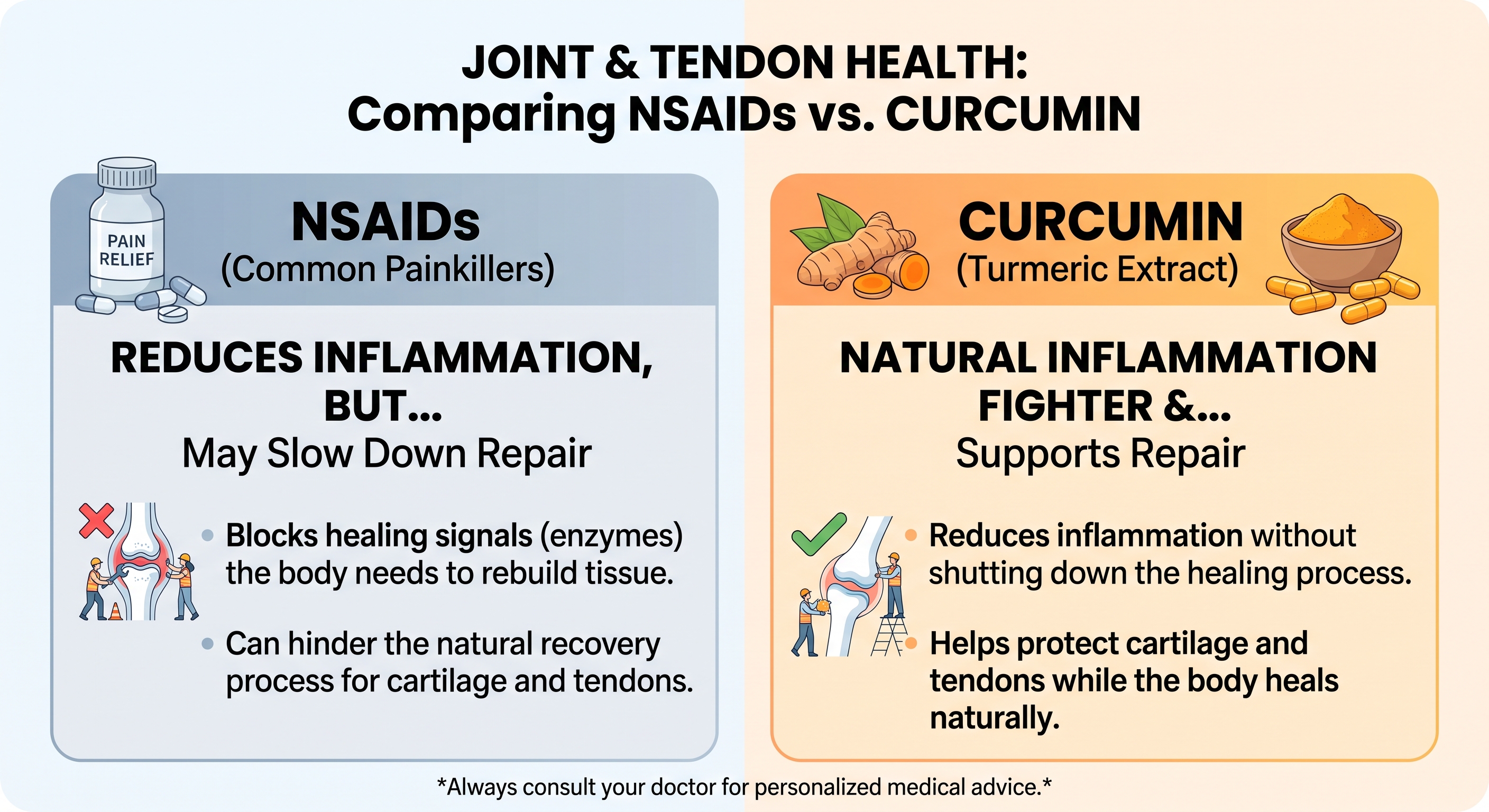
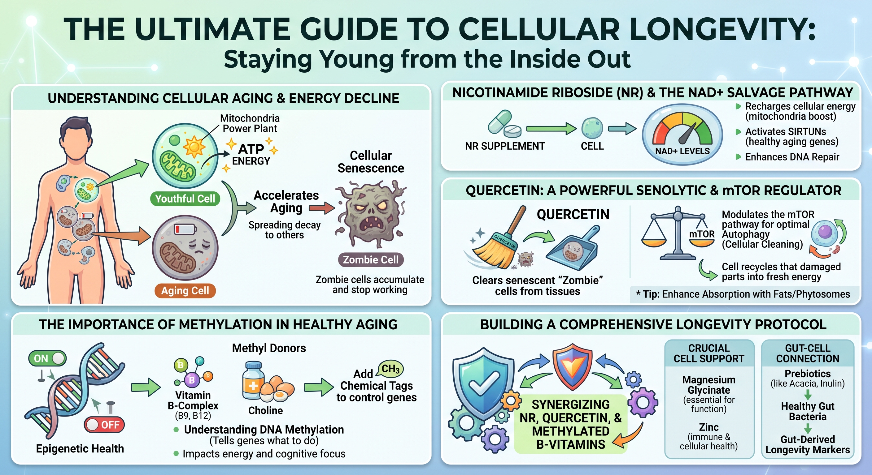
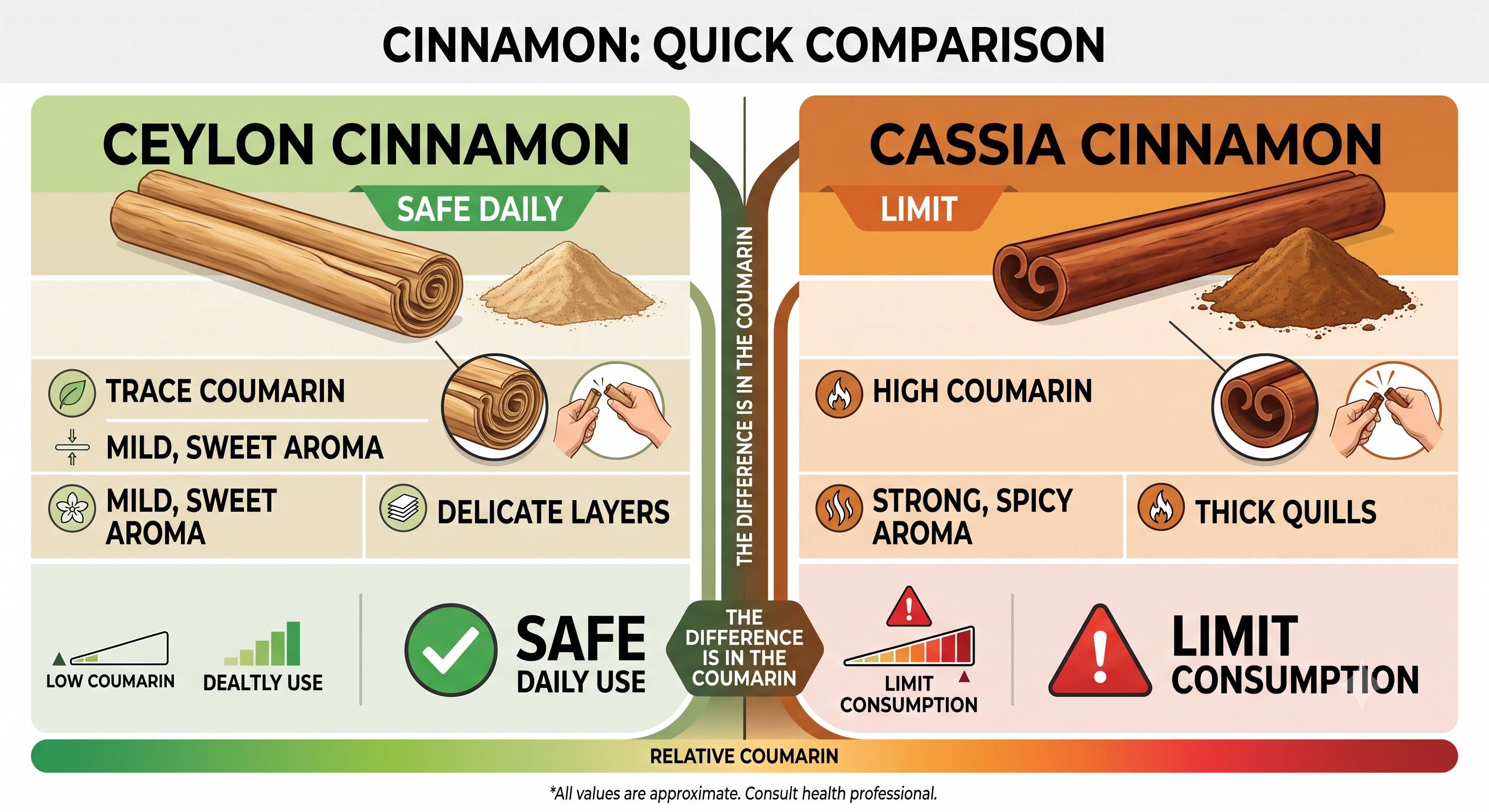

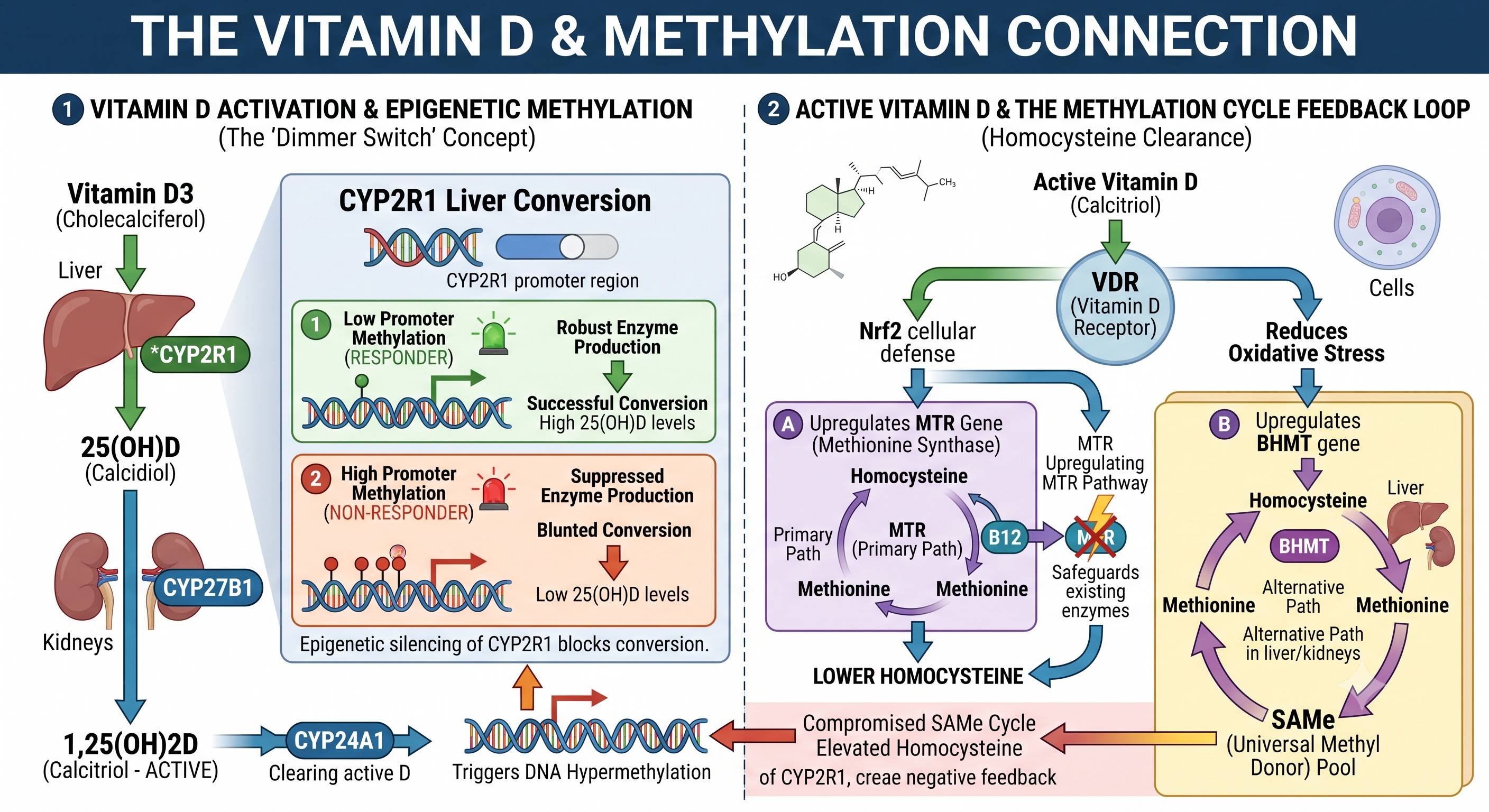
 The liver is a vital organ in the body that performs many essential functions. It can be damaged by a variety of factors, including disease, alcohol abuse, and exposure to toxic chemicals. Once the liver is damaged, can it be repaired? In this article, we will explore the answer to that question. We will also discuss the various ways that the liver can be damaged and how those damages can be repaired.
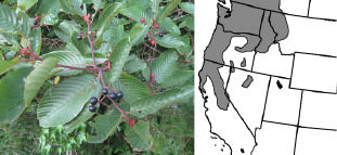
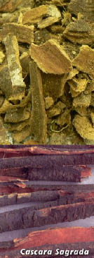 system. More research is needed before it will be recommended for treatment, but so far, the findings have been promising.
  Royal jelly is considered to be the treasure of the beehive. It has the ability to promote longevity by helping to maintain healthy, beauty, and youth. This natural food is extremely potent and highly nutritional. Royal jelly is an incredibly rich, creamy, opalescent white liquid. It is produced by the worker bees specifically for the nourishment and cultivation of the queen bee. This fantastic material converts a common honeybee into a queen bee, extending the bee’s lifetime from six weeks to five years. This substance is full of natural hormones and B-vitamins. Additionally, it contains a variety of seventeen amino acids. Eight of these amino acids are essential. Royal jelly is particularly rich in cystine, lysine, and arginine and is also composed of 16.1 percent aspartic acid, which is crucial for proper tissue growth and regeneration.
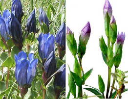 The gentian plant is an herbaceous perennial with fleshy root and fat hollow stems. The leaves of the plant are ovate and pleated, while the flowers are yellow. The root of the plant often smells aromatic and tastes sweet at first and then bitter.
 The horseradish plant is a perennial plant that is part of the Brassicaceae family, which includes mustard, wasabi, broccoli, and cabbages. Native to southeastern Europe and western Asia, the plant is popular around the world today. The horseradish plant grows up to five feet tall and is mainly cultivated for its large, white, tapered root. The intact horseradish root has hardly any aroma. However, when cut or grated, enzymes from the damaged plant cells break down to produce allylisothiocyanate, which often irritates the sinuses and eyes. Once grated, if the plant is not mixed with vinegar or used immediately, the root darkens and loses its pungency. It quickly becomes unpleasantly bitter when exposed to air and heat.
  This herb helps promote stomach secretions to aid in digestion. Horseradish can be used as a compress for neuralgia, stiffness, and pain in the back of the neck. Additionally, this herb can be used as a parasiticide.
  Goldenseal was used by the Native Americans as a tonic, for sore throats, eye infections, ulcers, and even arrow wounds. It was also used as an insect repellant and pesticide for crops. When boiled in water, it was used externally for skin conditions. The dried root of the